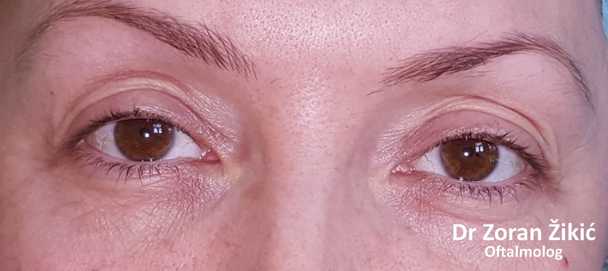
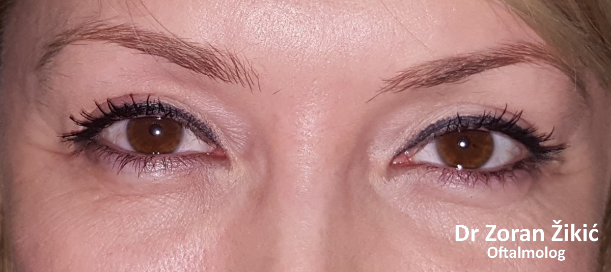
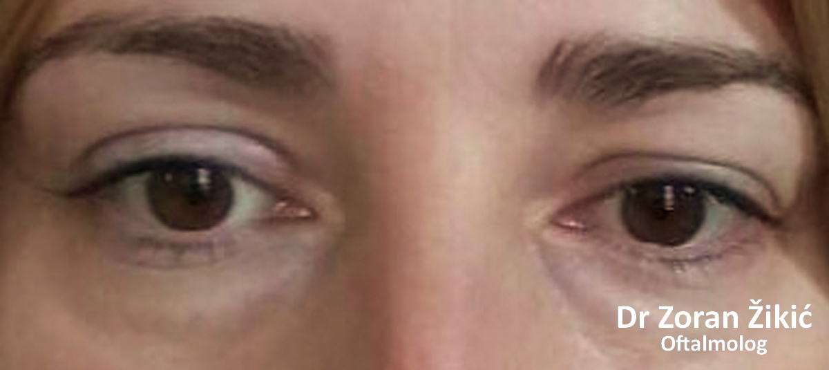
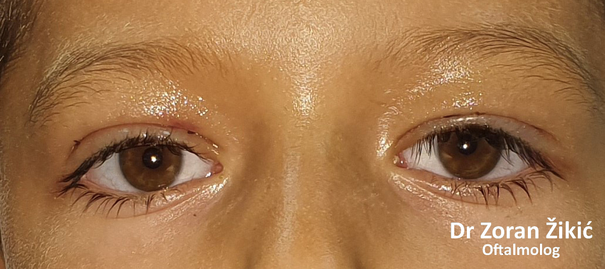
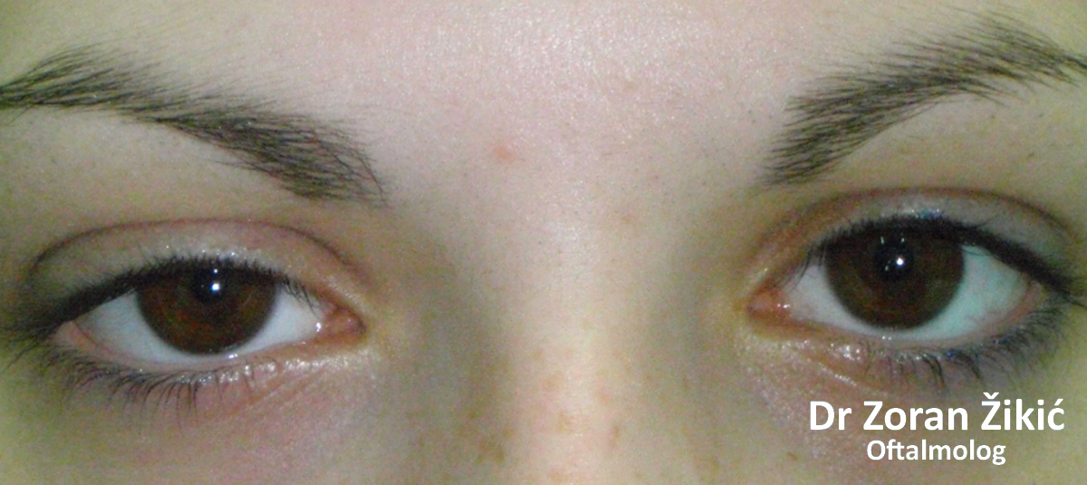
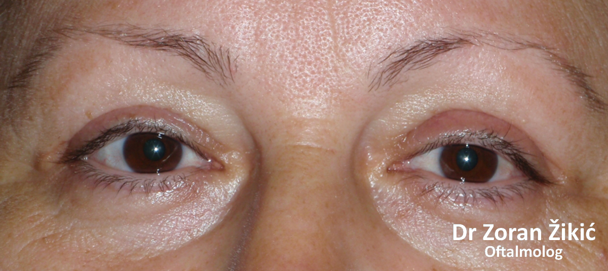
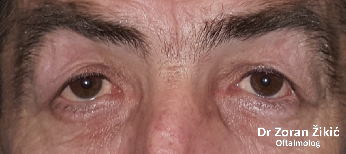
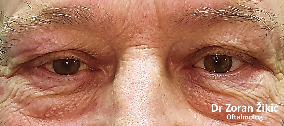
Lower lid bags and deep „tear trough“ (junction between lid and cheek).
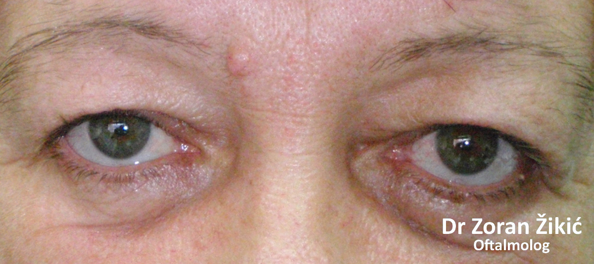
One month after lower lid blepharoplasty with fat repositioning and skin tightening.
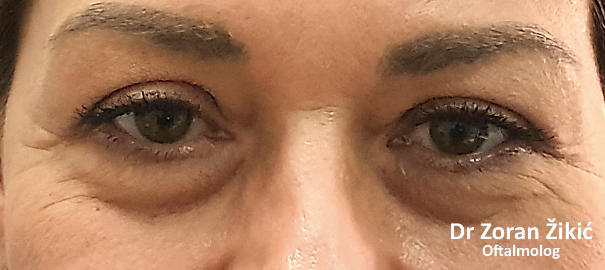
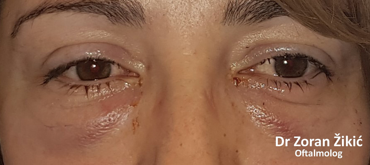
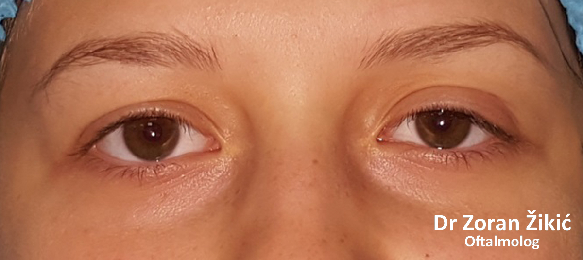
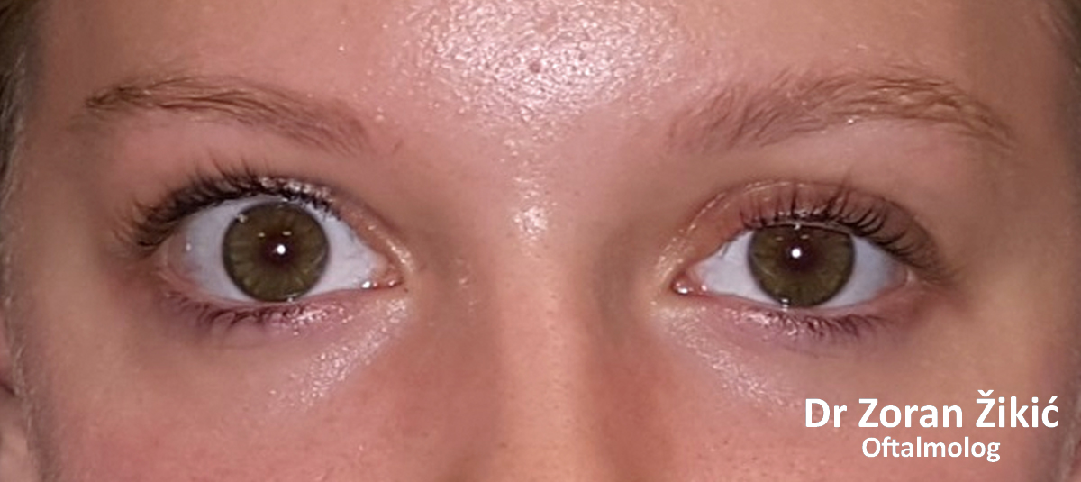
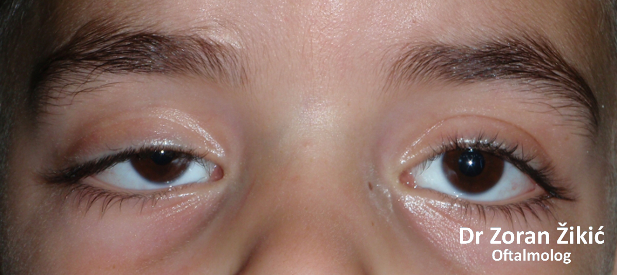
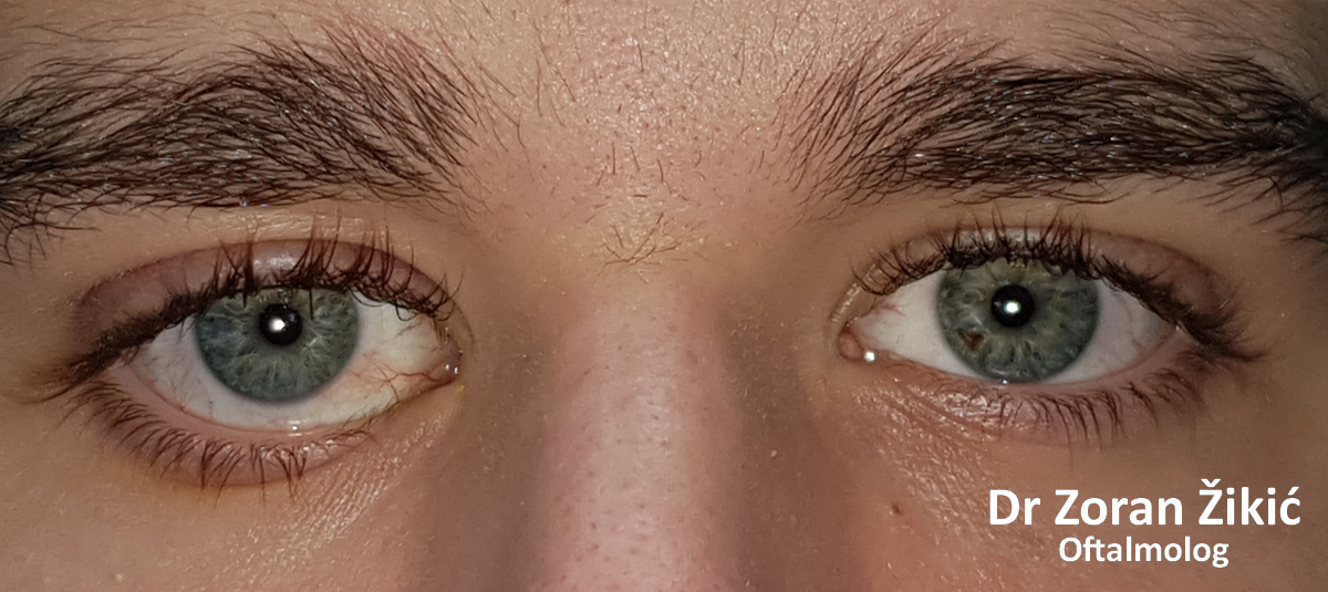
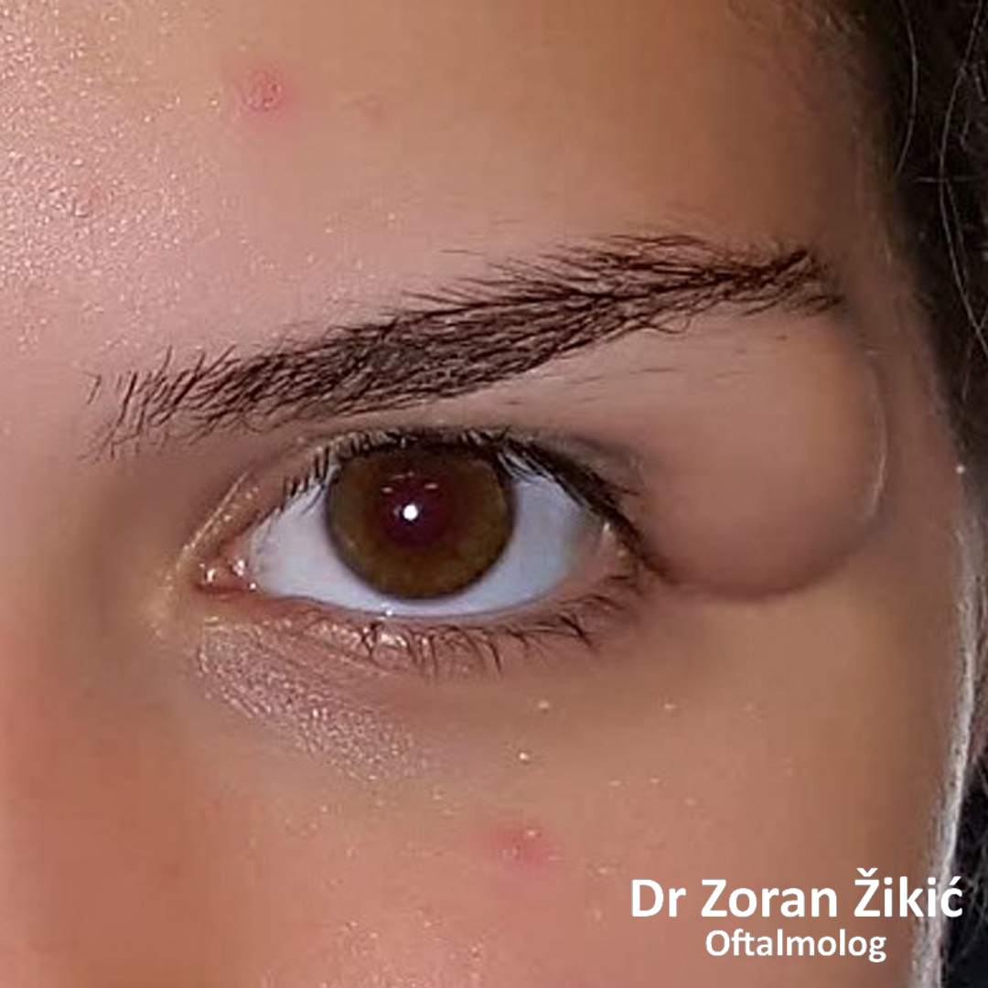
Under eye bags, with good skin tone (selfie).
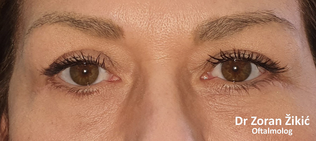
One day after lower lid fat reduction (partial removal)m without skin incision (transconjunctival blepharoplasty).
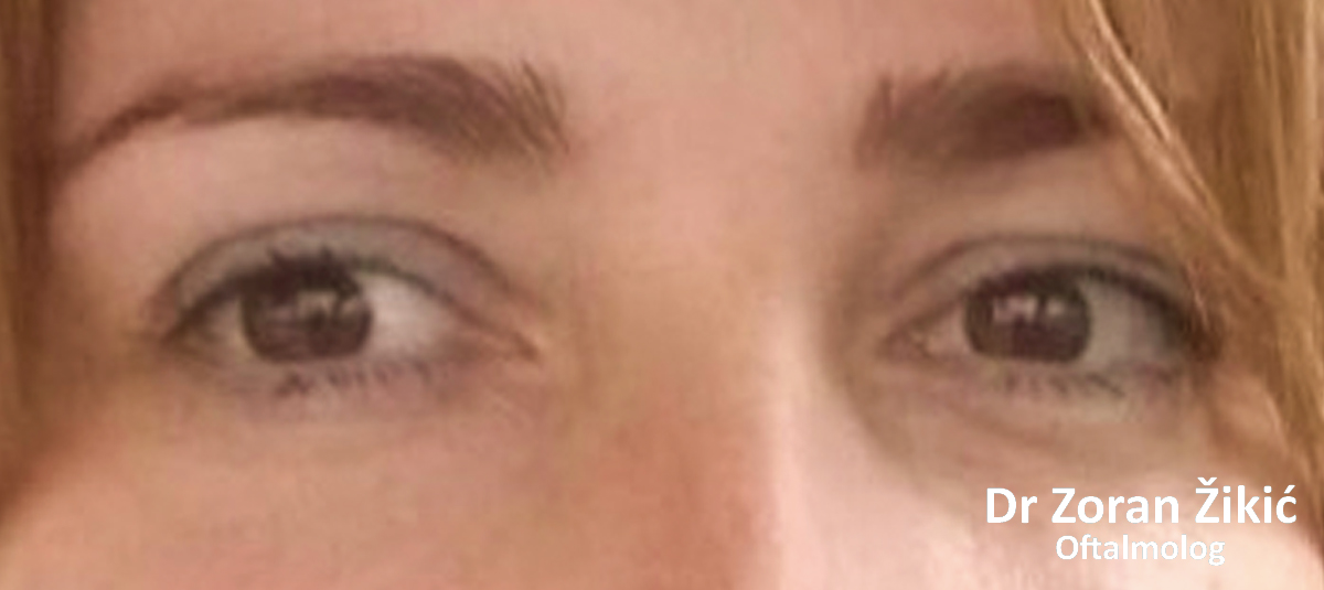
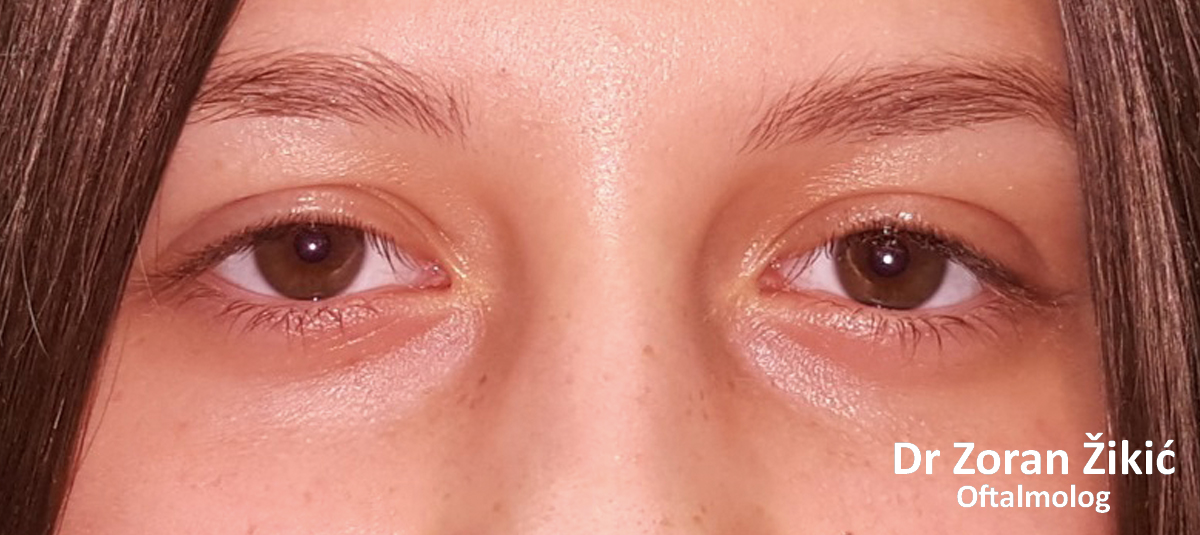
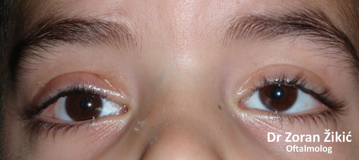
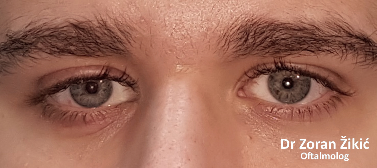
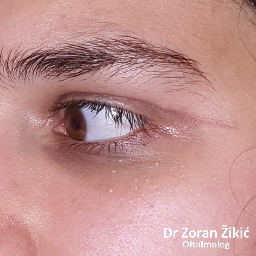
One month after surgery (selfie).
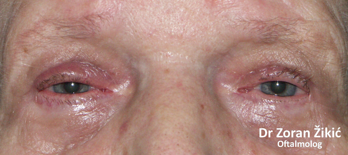
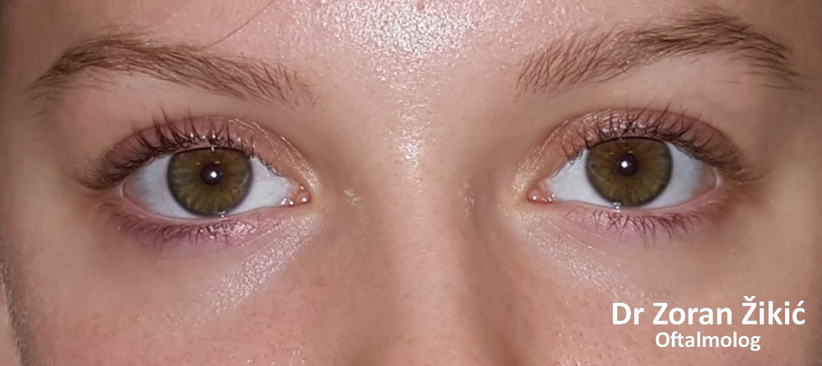
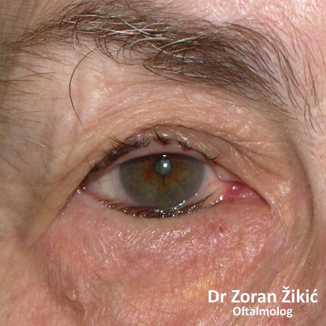
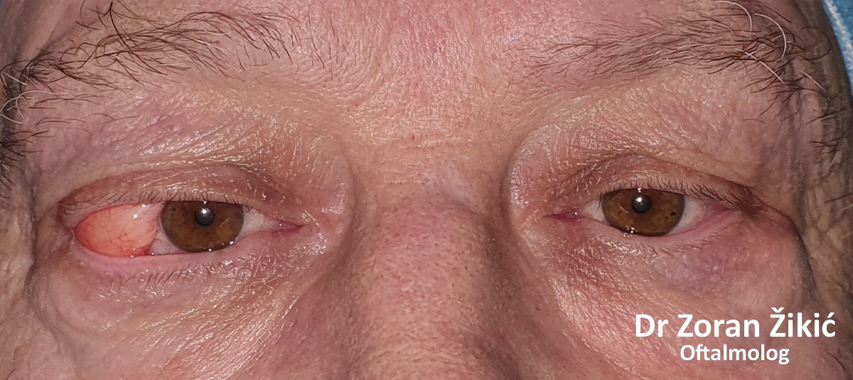
Prominent lower lid bags with stretched and lax skin.
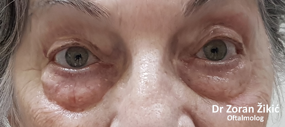
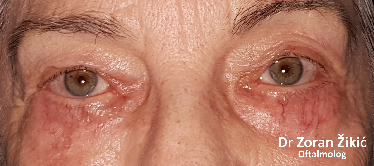
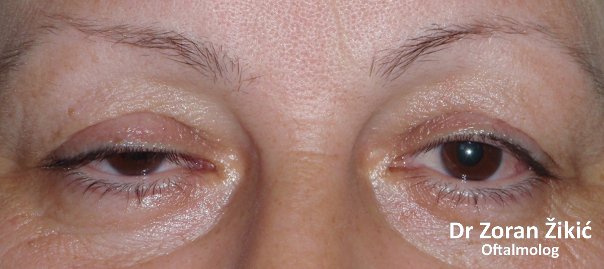
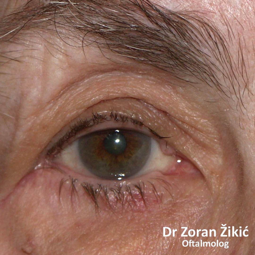
One month after lower lid blepharoplasty with fat repositioning and skin tightening. The blood vessels are visible because of very thin skin.
FUNCTIONAL EYELID SURGERIES
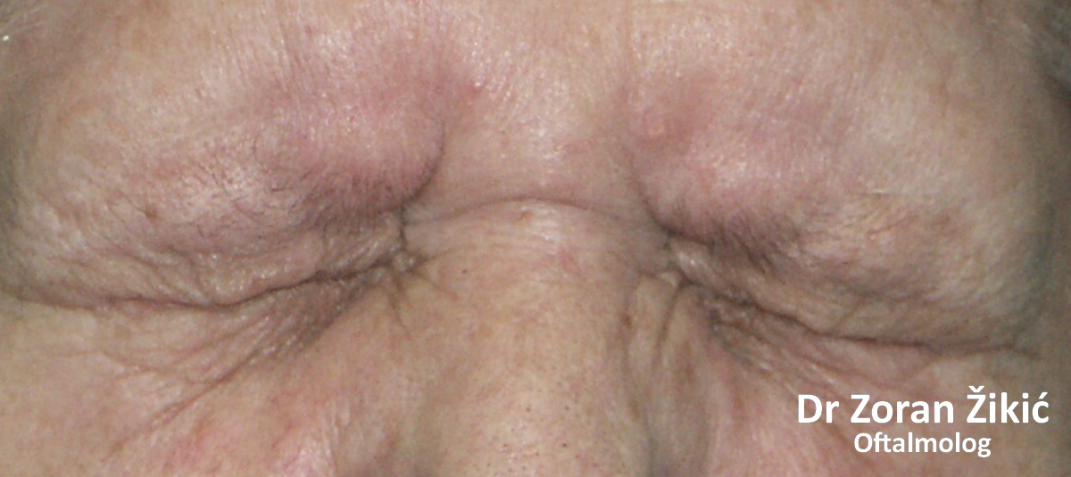
Excessive stretching of the skin (dermatochalasis) combined with lymphostasis in the eyelids.
After surgery.
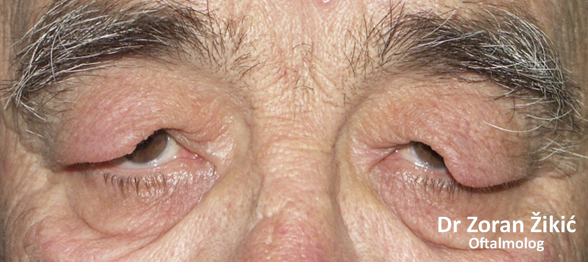
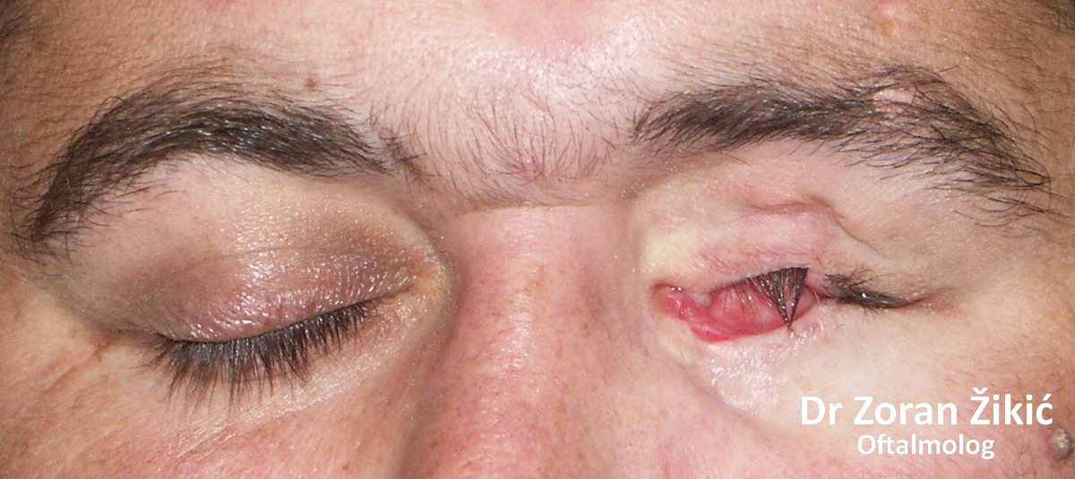
Inabbility of voluntary opening of the eyes, due to cramping of the eyelid muscles (blepharospasm). The patient did not react to treatment with botulinum toxin injections.
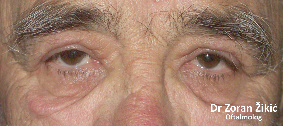
After surgical treatment.
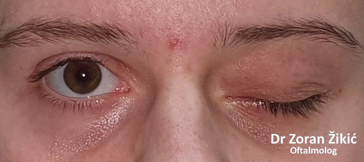
DROOPING UPPER LID (PTOSIS)
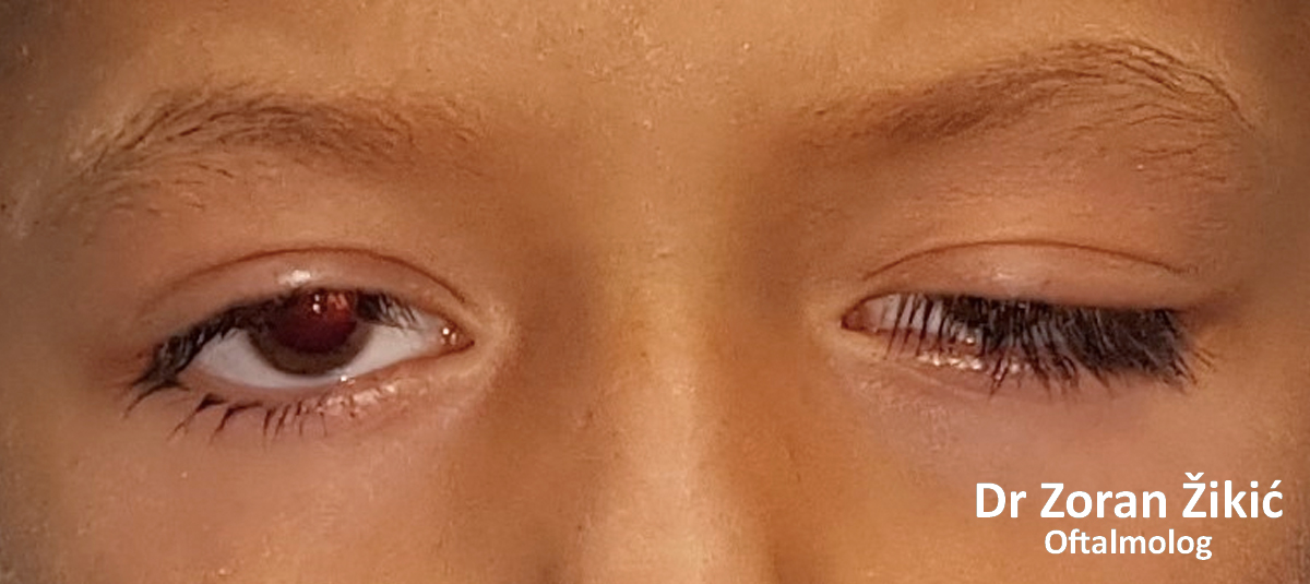
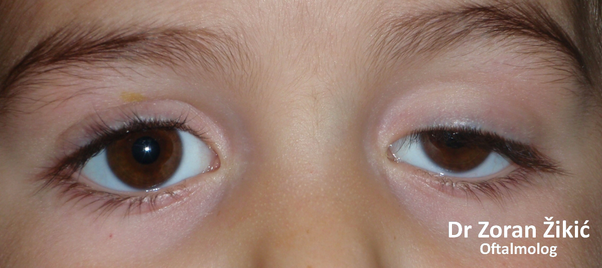
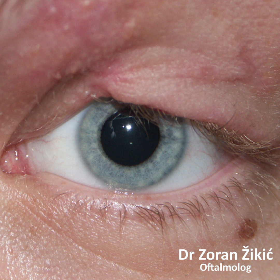
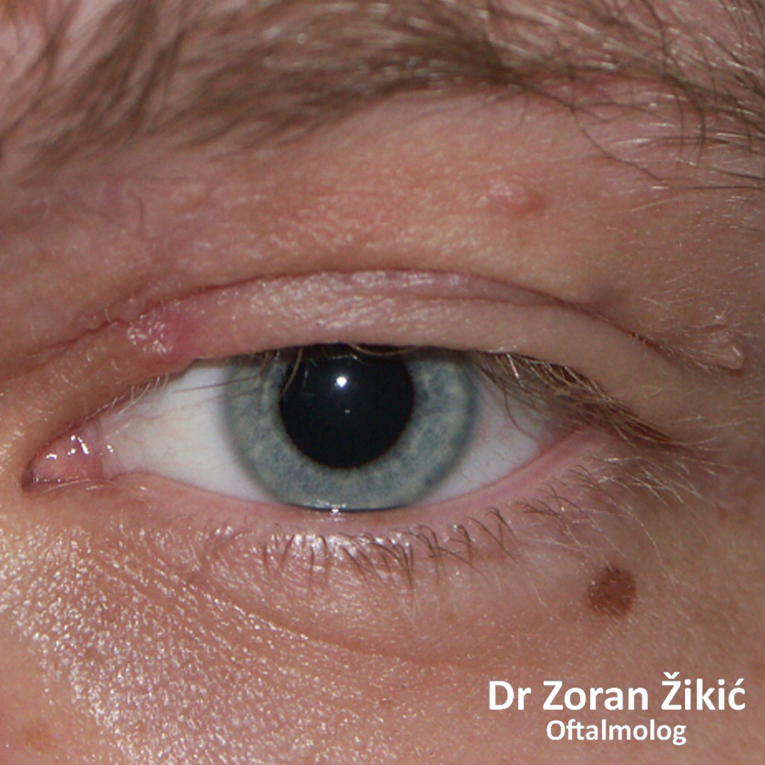
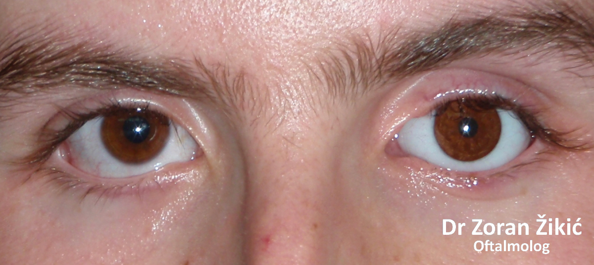
A discreet congenital ptosis on the left upper lid.
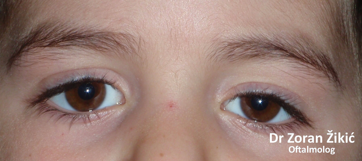
One month after transconjunctival (no skin inscision) correction of ptosis.
Congenital asymetrical ptosis of both upper lids.
Two weeks after ptosis corrcetion by levator resection on the right side, and by direct frontalis (forehead) muscle fixation on the left side.
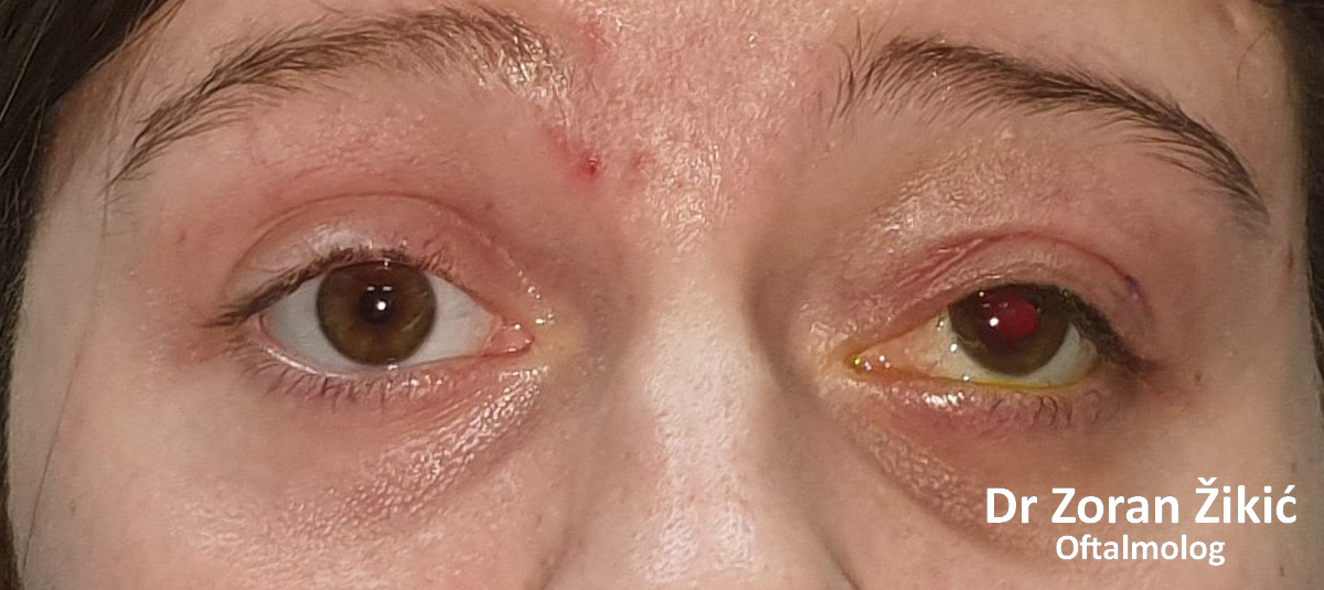
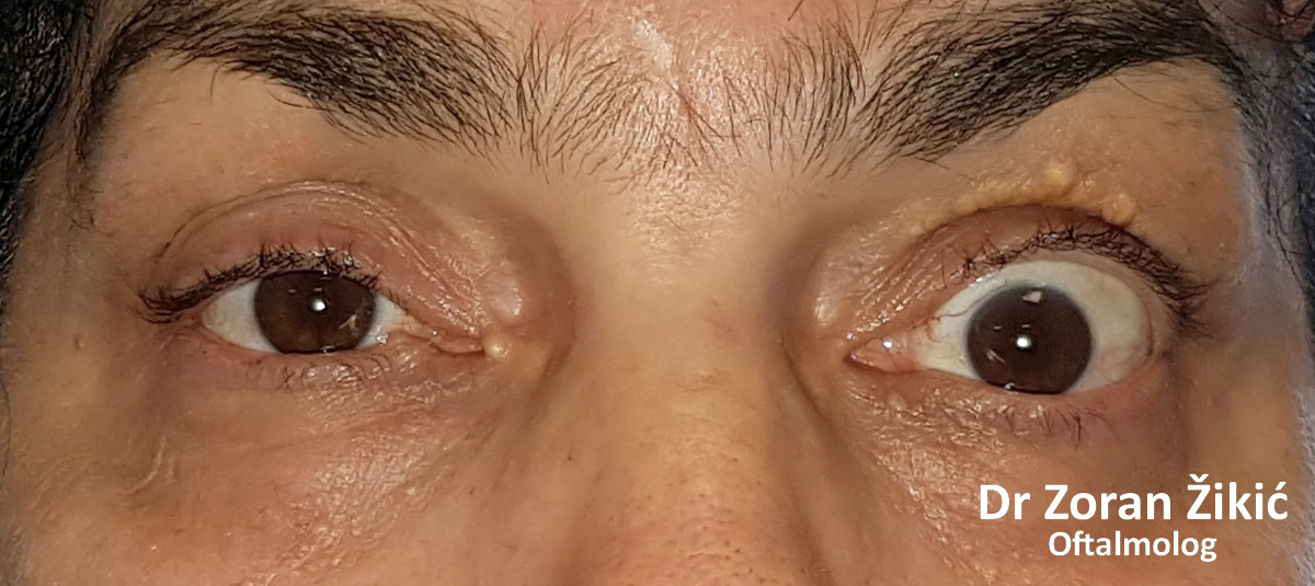
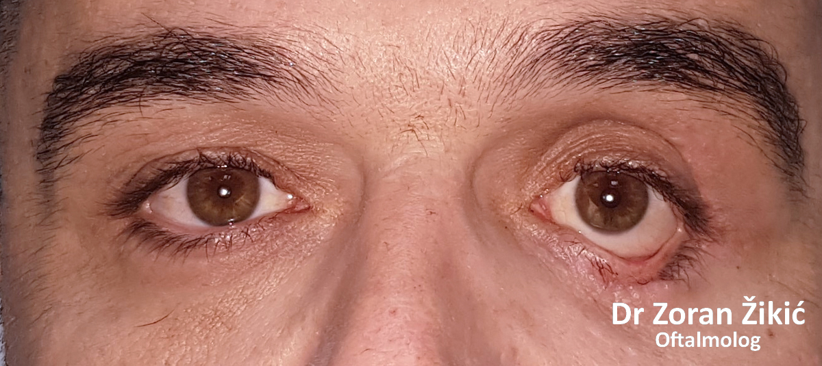
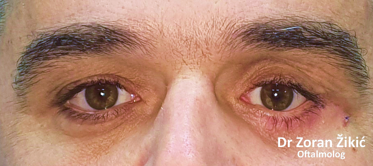
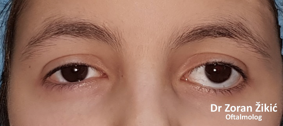
Ptosis (droopy upper lid) on the left side, with compensatory retraction On the right side.
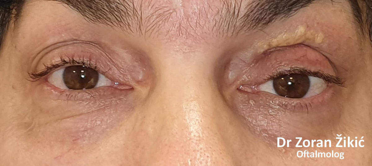
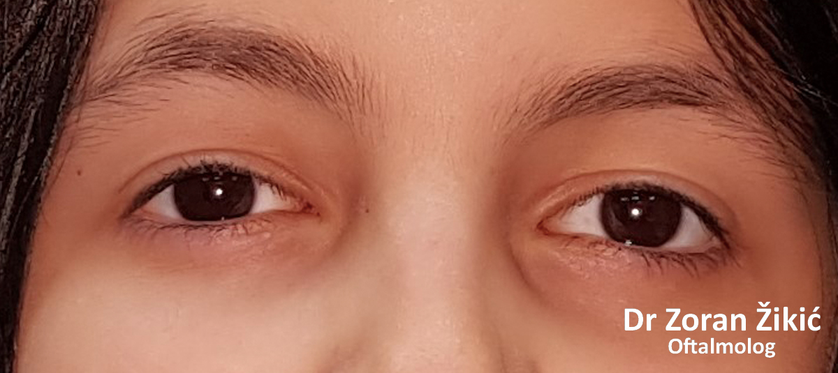
After transconjunctival (no skin inscision) correction of ptosis.
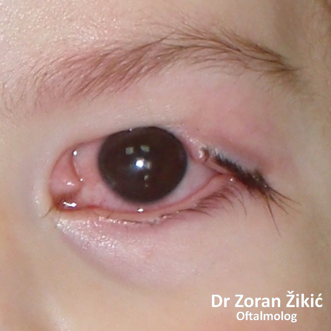
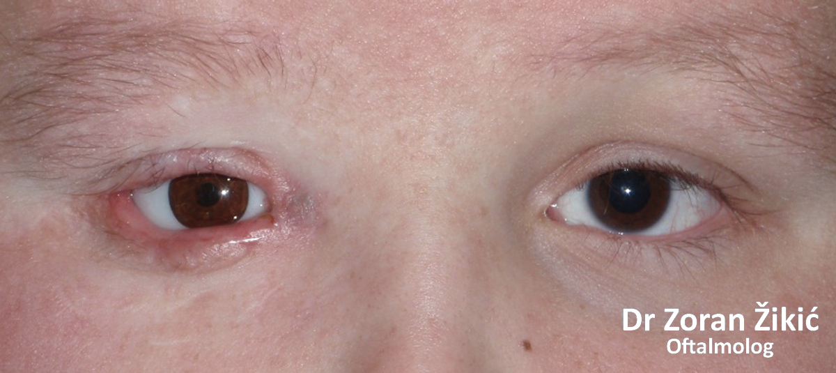
Complete ptosis of the left upper lid, fixated (immobile) left eye and wide pupil, due to oculomotorius or 3rd nerve palsy (paralysis).
One month after carefull sub-total correction, by direct fixation to the frontalis (forehead) muscle.
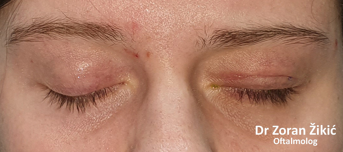
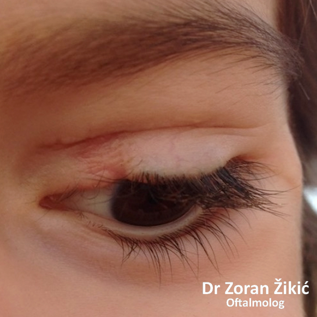
Good closure of left eye.
Congenital drooping (ptosis) of the left upper lid.
After correction with levator muscle resection (shortening)..
Congenital drooping (ptosis) of the right upper lid.
After correction with levator muscle resection (shortening)..
Congenital ptosis of the right upper lid.
After correction by frontalis suspension with a silicone sling.
Acquired drooping (ptosis) of the right upper lid.
After correction by reinsertion of the levator aponeurosis.
Acquired drooping (ptosis) of the left upper lid.
After correction by reinsertion of the levator aponeurosis.
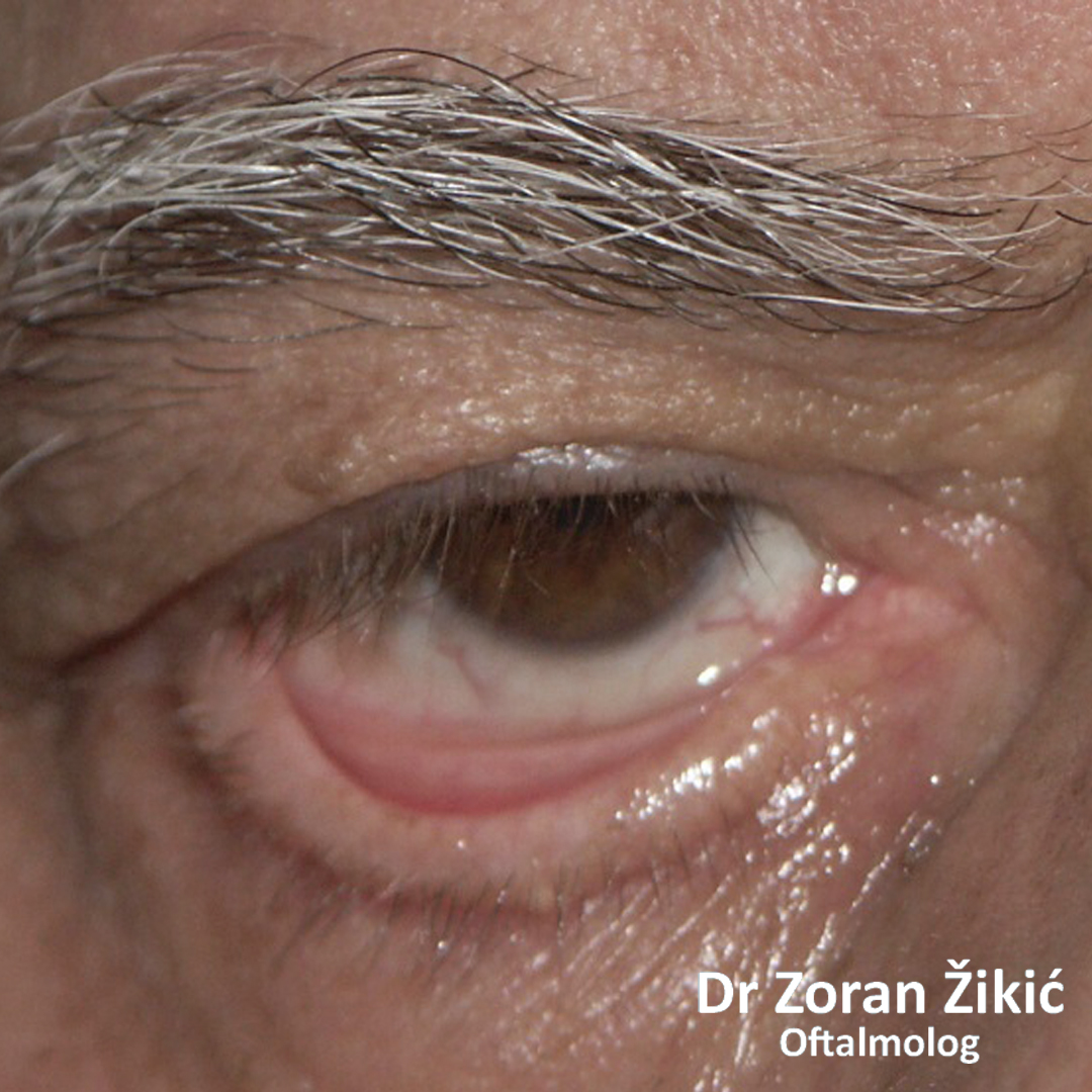
RETRACTION OF THE EYELID
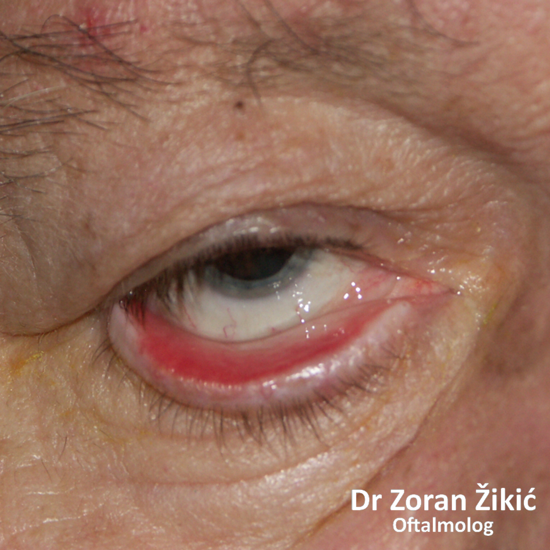
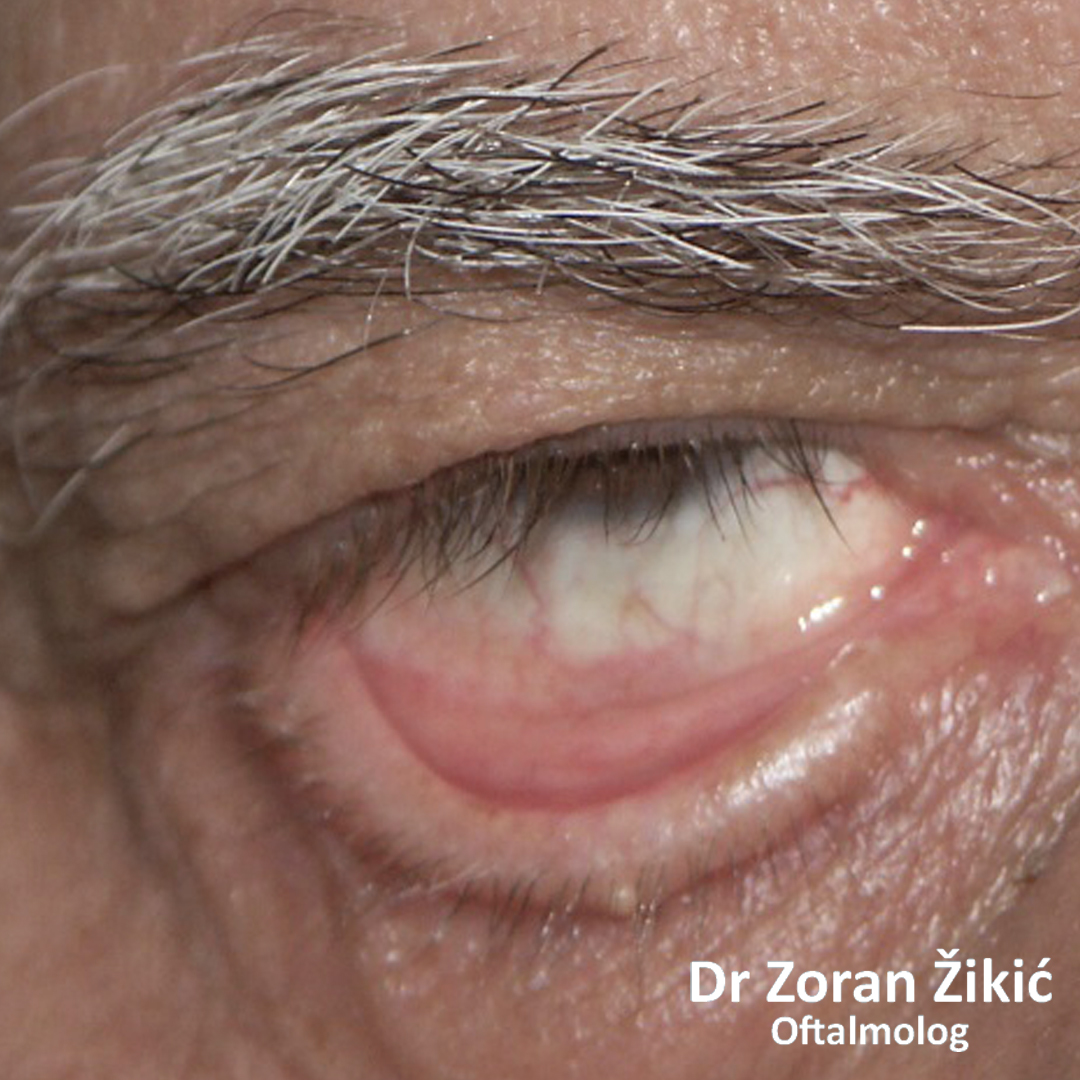
Retraction of the right lower lid.
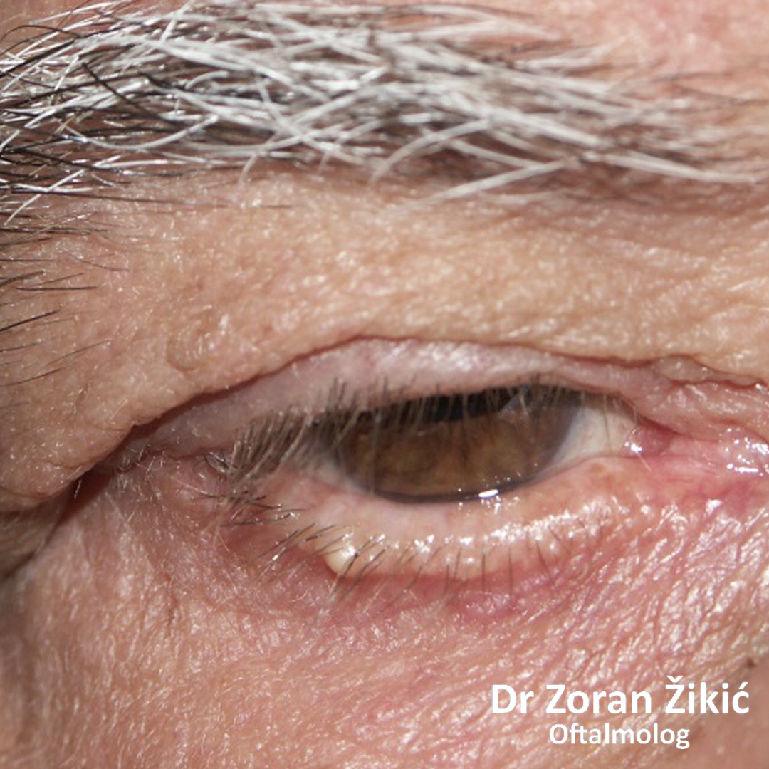
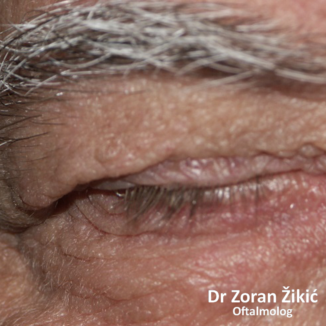
After correction of retraction with hard pallate mucosa transplantation.
Retraction of the left upper lid, due to endocrine orbitopathy.
After correction of retraction by recession of the upper lid retractors (elevator muscles).
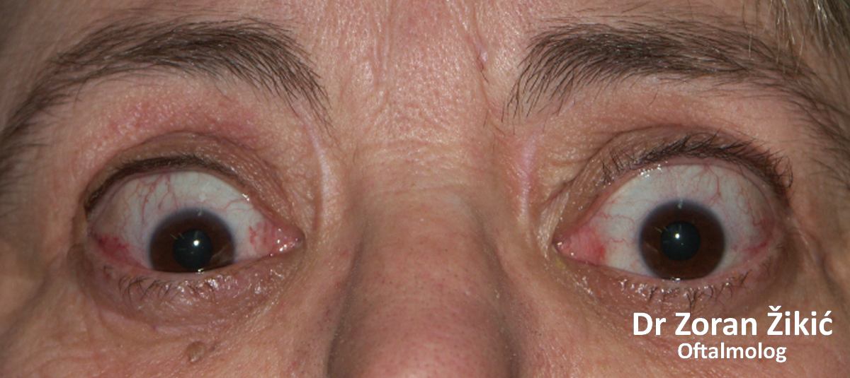
Retraction of the upper eyelids in Graves diseas.
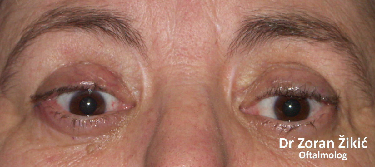
After surgical correction.
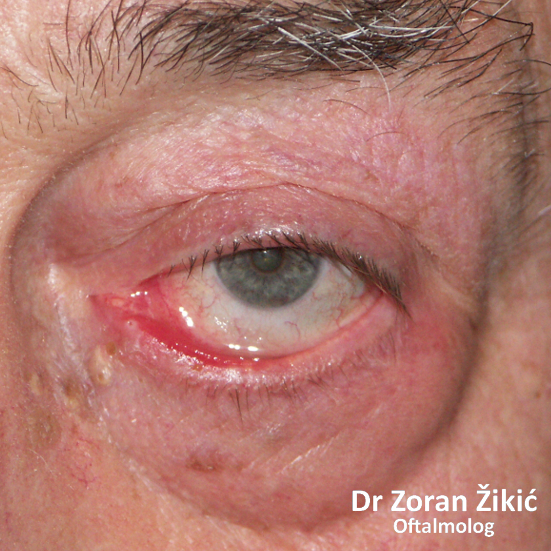
EVERTED (ECTROPION) AND INVERTED (ENTROPION) EYELID
Eversion (ectropion) of the lower lid.
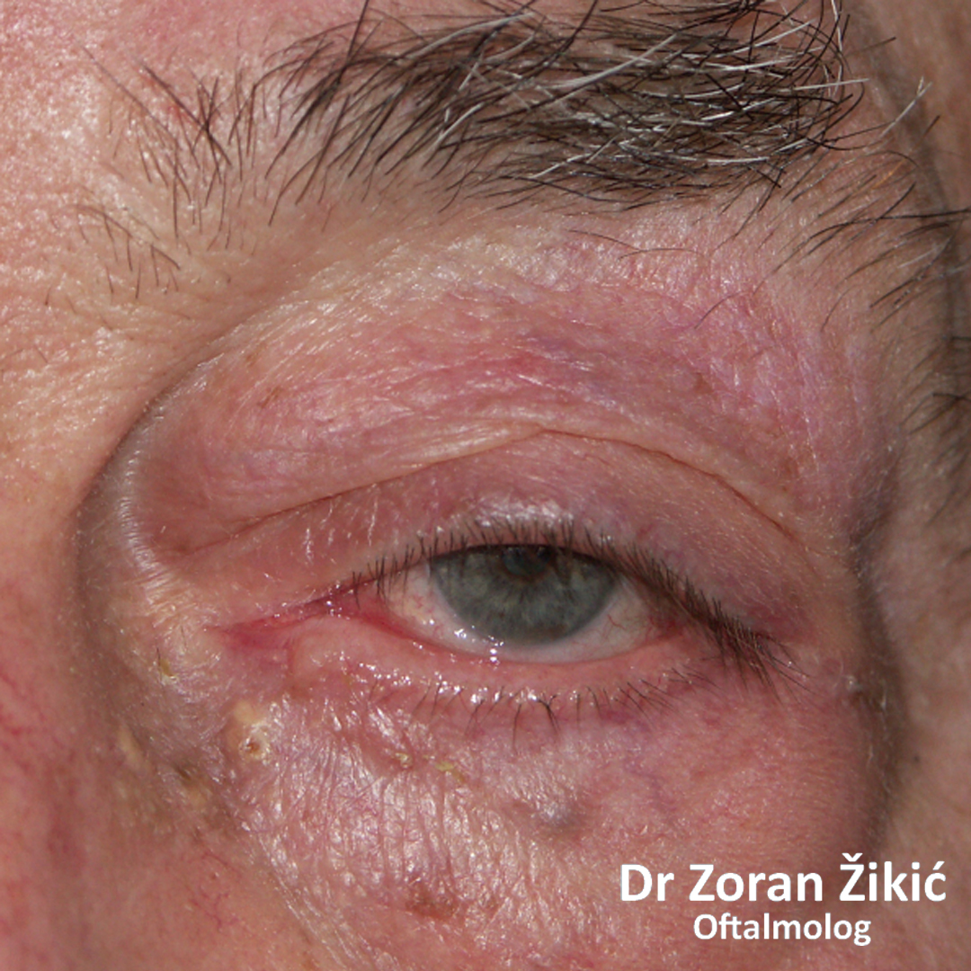
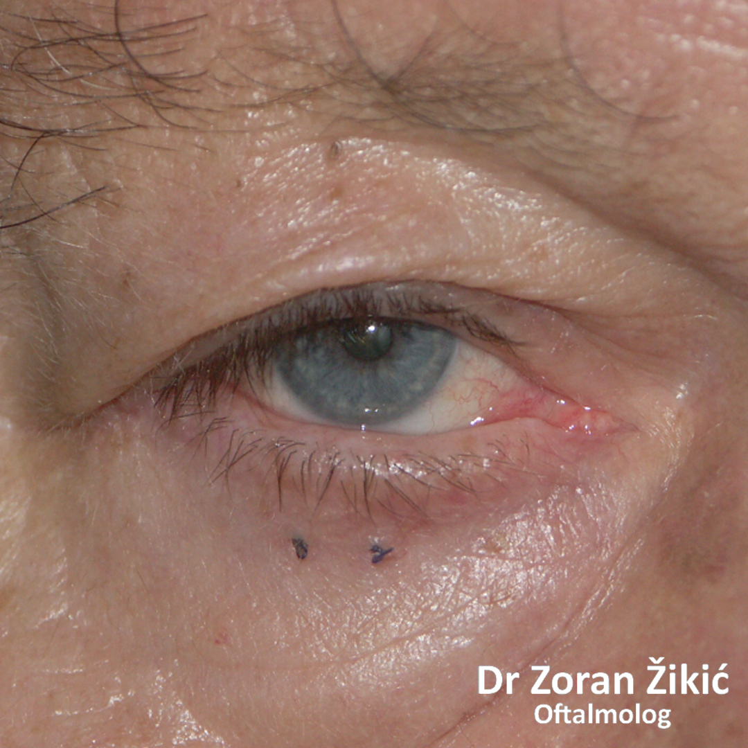
After operation.
Eversion (ectropion) of the whole lower lid.
2 weeks after the operation (the sutures on the lower lid are self resorbable and fall out after about one month).
Eversion (ectropion) of the lower lid with the inability of the closure of the eye, due to palsy of the facial nerve.
After surgery.
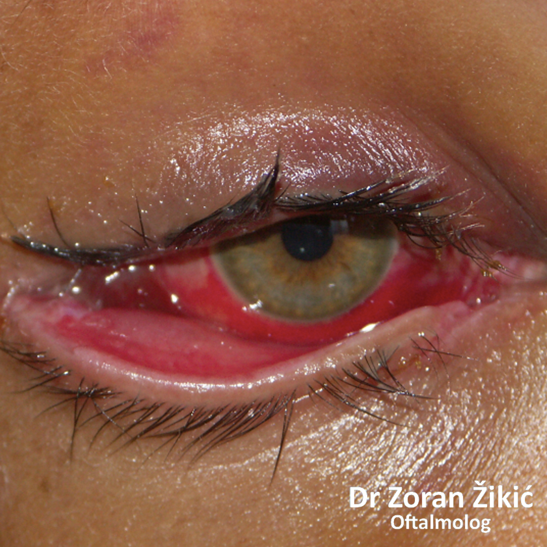
Inversion (entropion) of the lower lid, with iflammation of the eye due to eyelash abrasion.
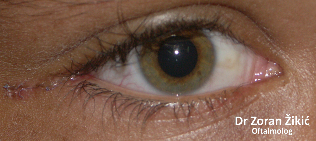
After operation.
POVREDE I OŽILJCI KAPAKA
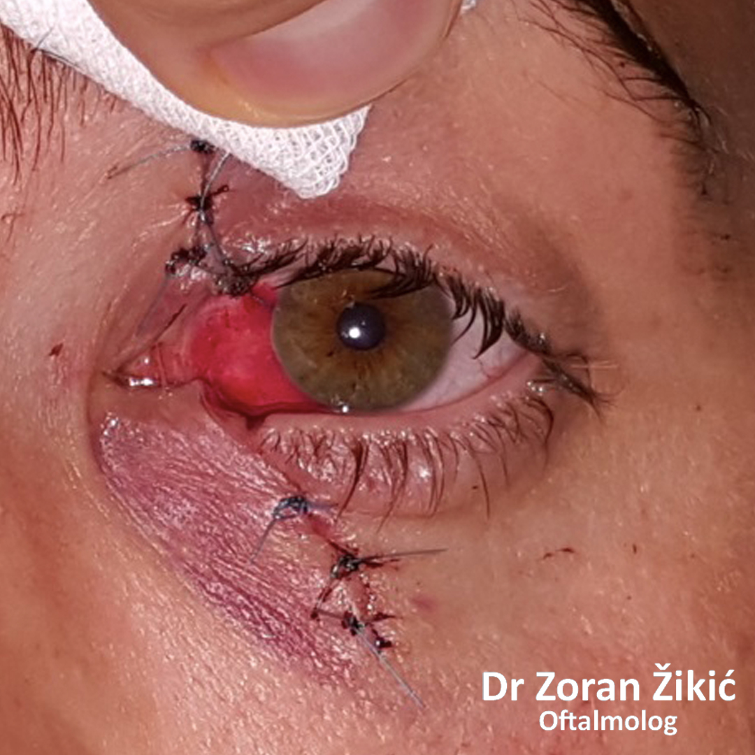
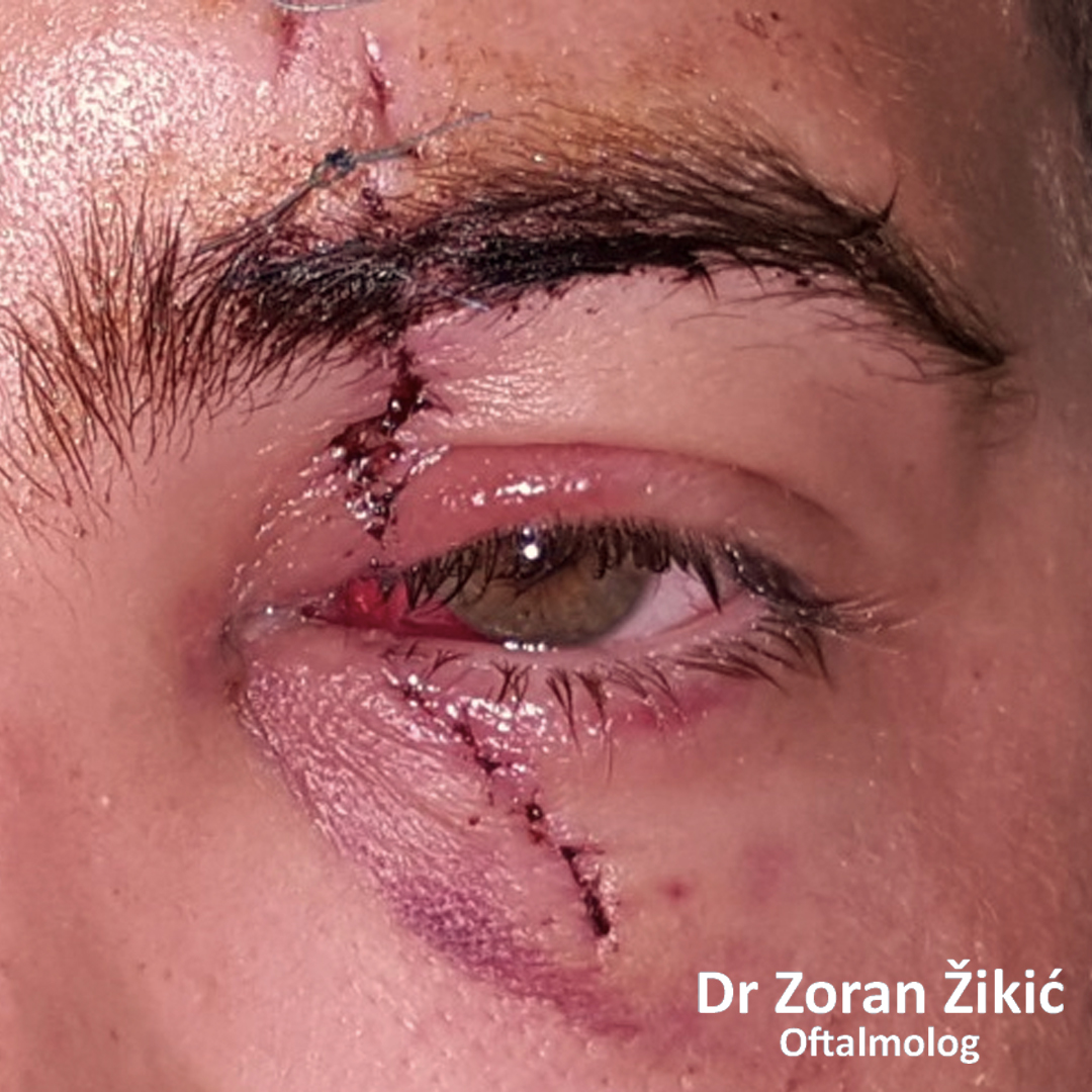
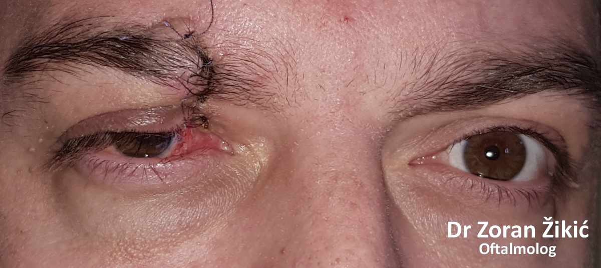
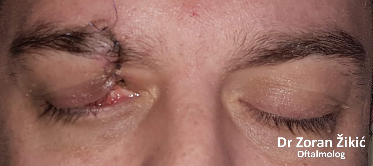
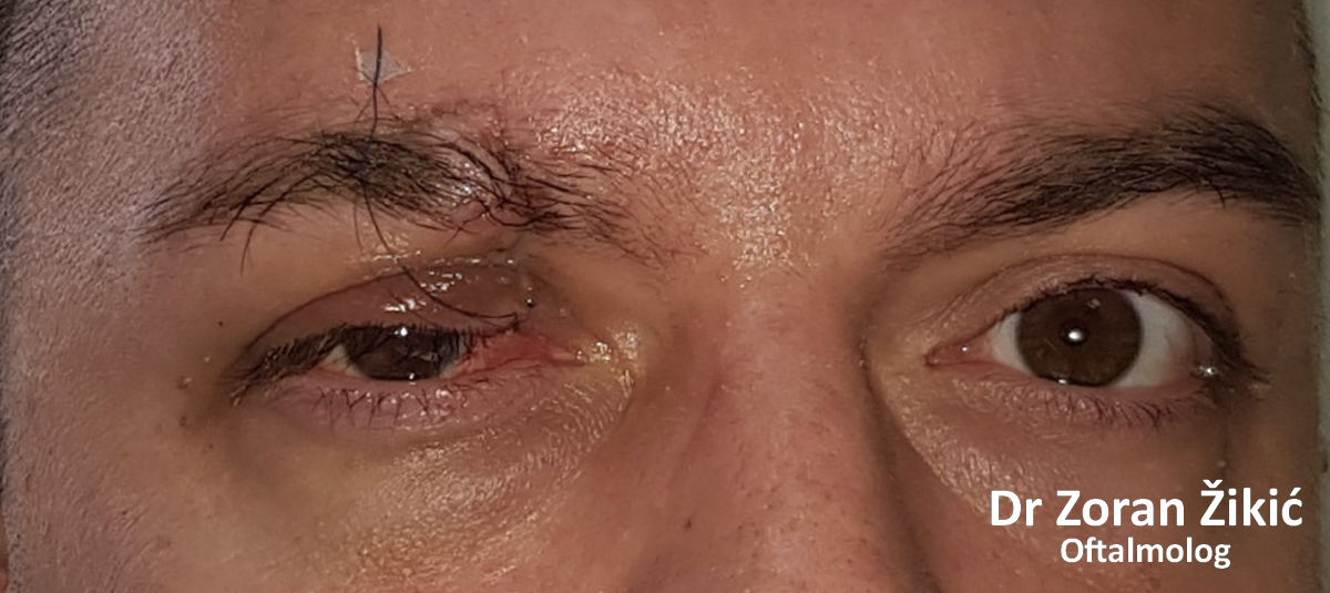
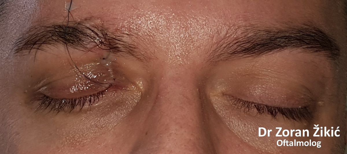
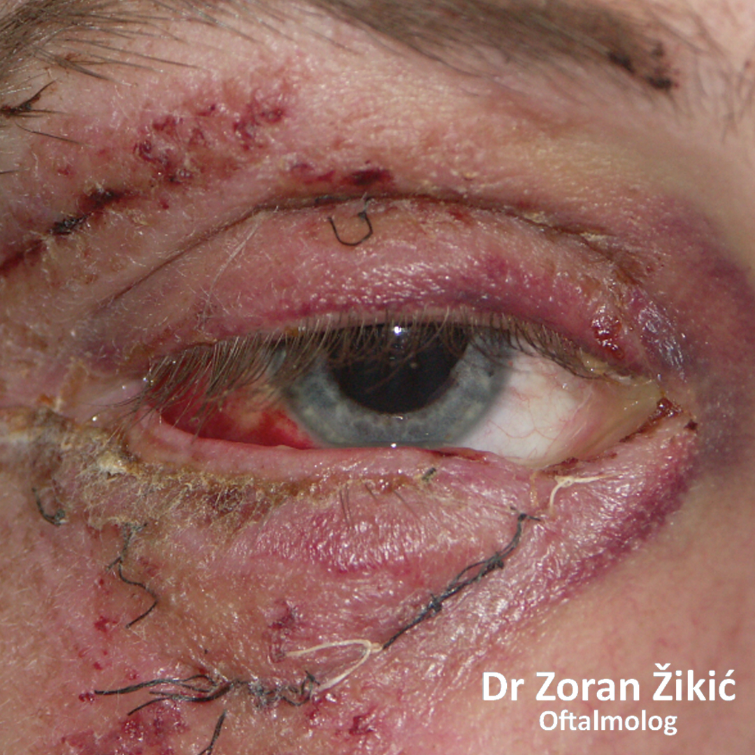
Laceration of both lids of the left eye, treated elsewhere. The patient complains of scratching and tearing of the left eye.
One week after secondary repair.
After primary repair of right upper lid laceration elsewhere. The patient complains of right eye scratching and incomplete closure of the right eye.
One week after secondary repair.
Cicatricial (scar induced) retraction of left lower lid, after fracture of the upper jaw bone (maxilla).
After two sequential corrective surgeries.
Defect of the upper lid (coloboma) after trauma.
After surgery.
Injury of the lower lid, after an inadequate repair elsewhere.
After reconstruction.
Injury of the lower lid..
After reconstruction.
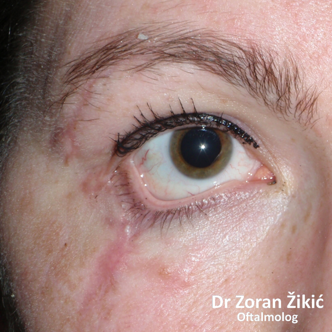
Retraction of the lower lid due to scarring (cicatriceal retraction) after trauma.
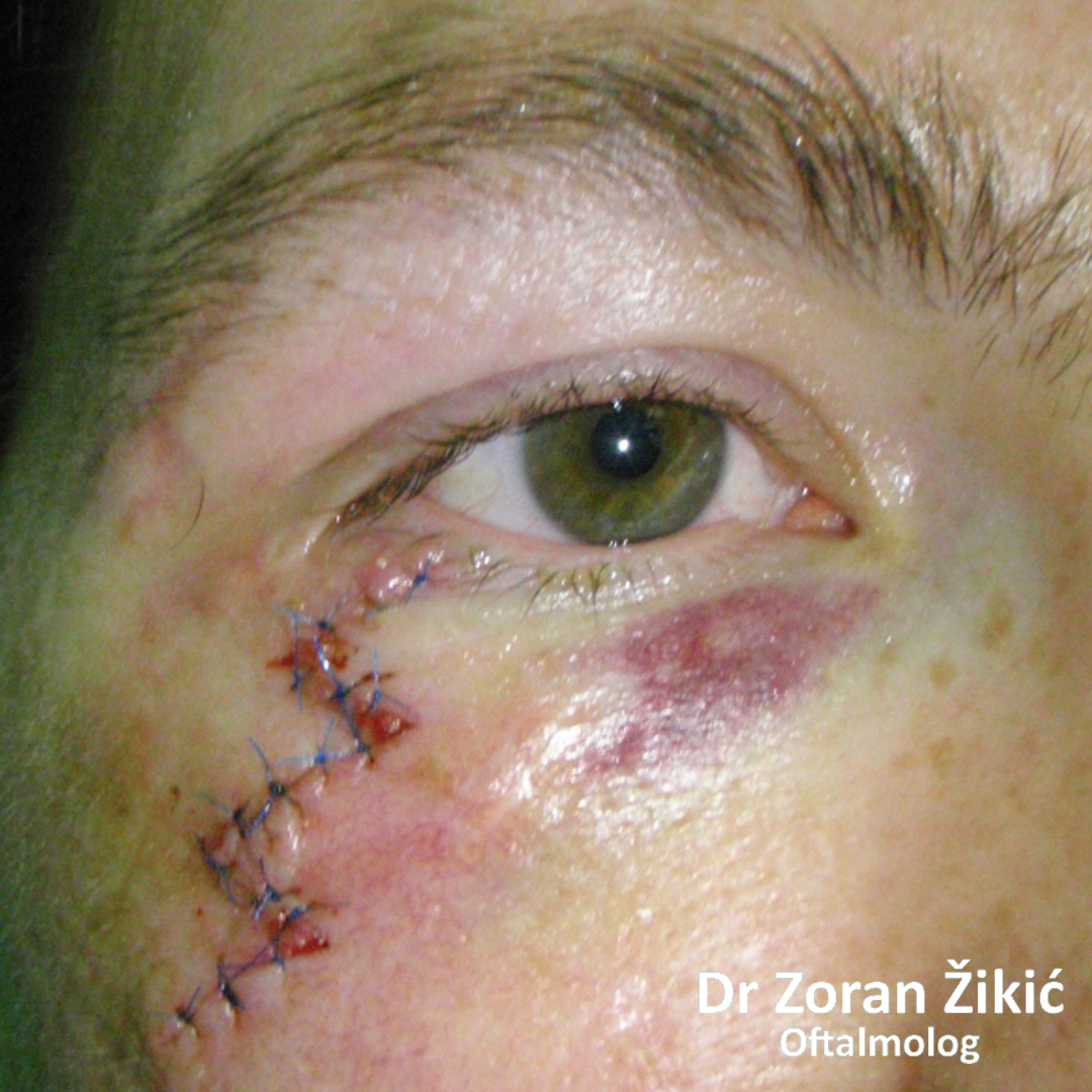
days after surgical correction (sutures to be removed).
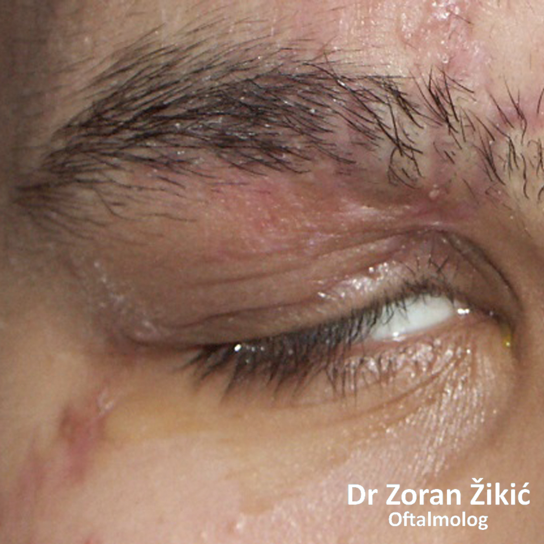
Inability to close the upper lid (lagophthalmos), due to scarring after trauma.
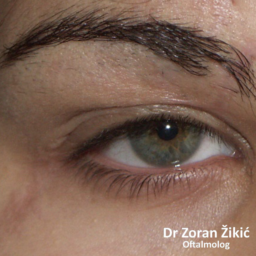
After surgical reconstruction..
TUMORI OKA, KAPAKA I PERIOKULARNE REGIJE
Cystic enlargement of tear gland on both sides.
Confirmation of cysts by trans-illumination.
After surgical removal of cysts.
Cyst of the left upper lid.
After surgical removal of the cyst.
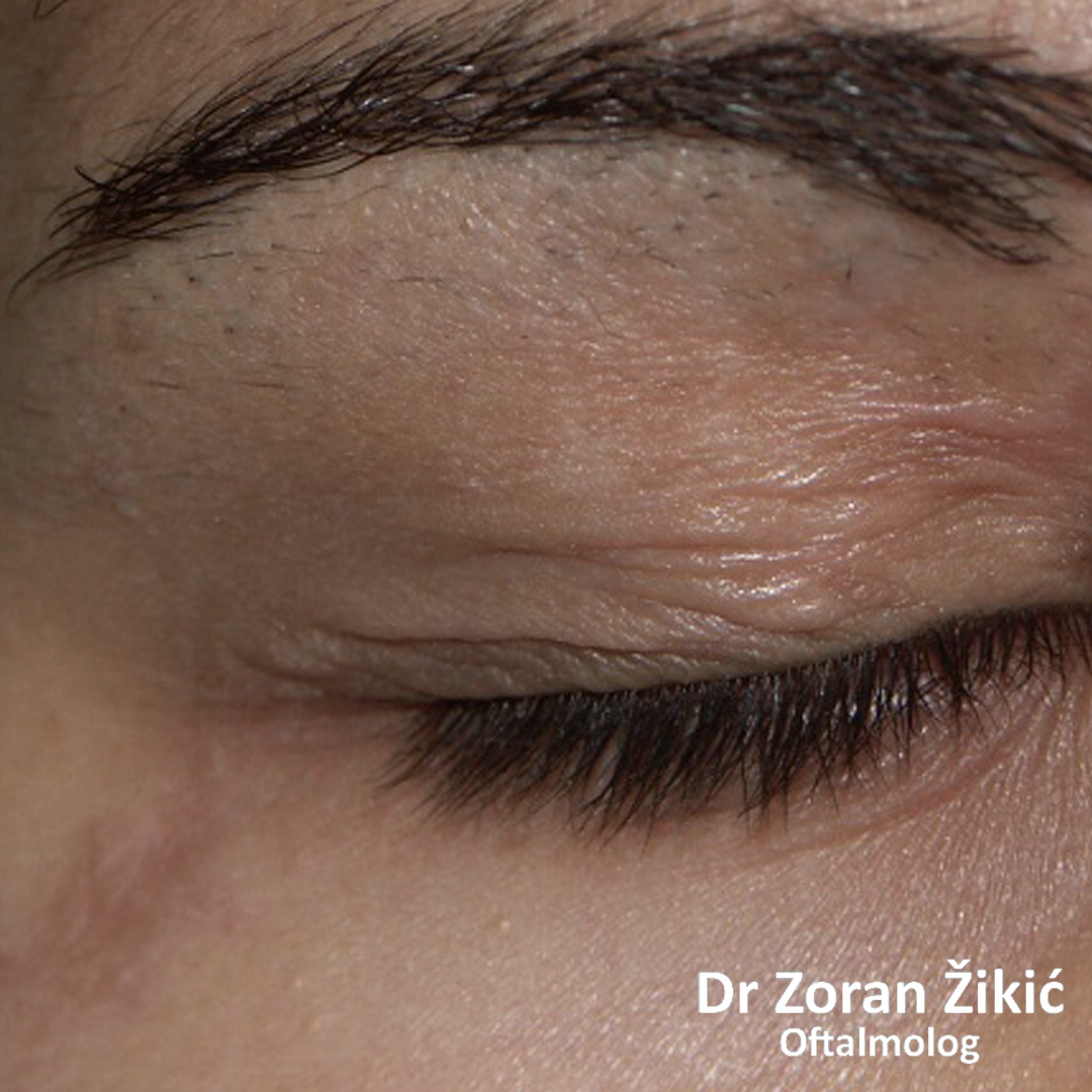
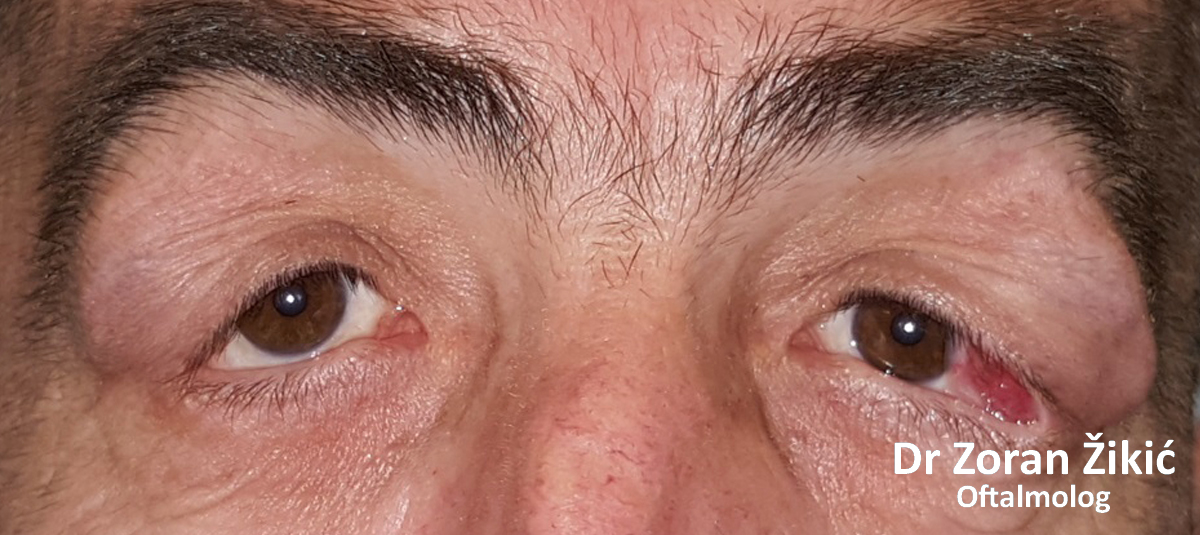
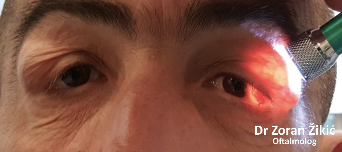
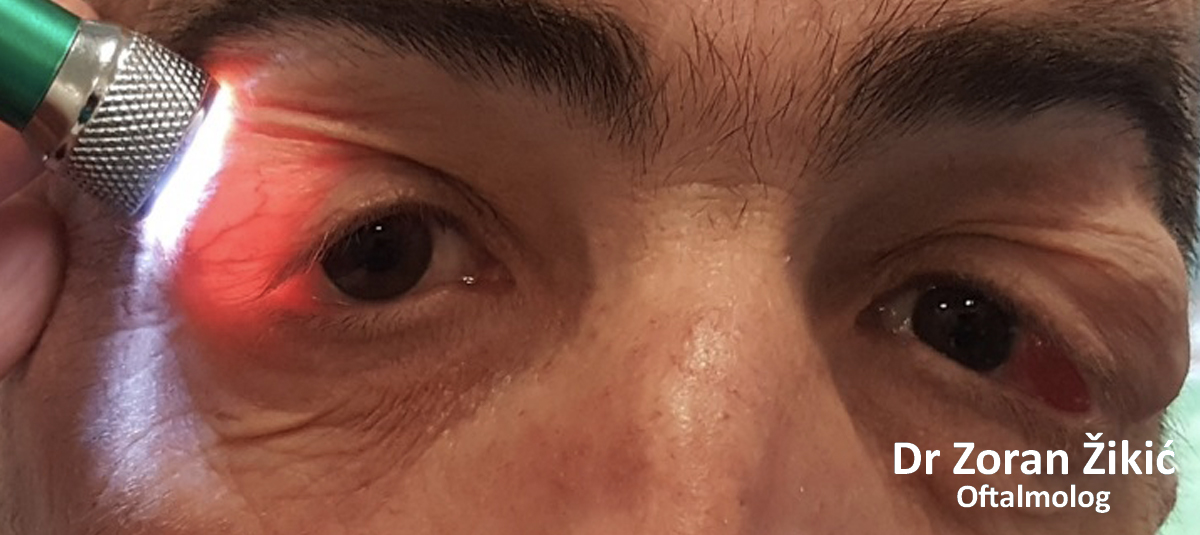
Prolapse or „herniation“ of orbital fat, under the eye mucosa (conjunctiva).
After micro-surgical removal of prolapsed fat and repair of the fibrous septa (Tenon-plasty).
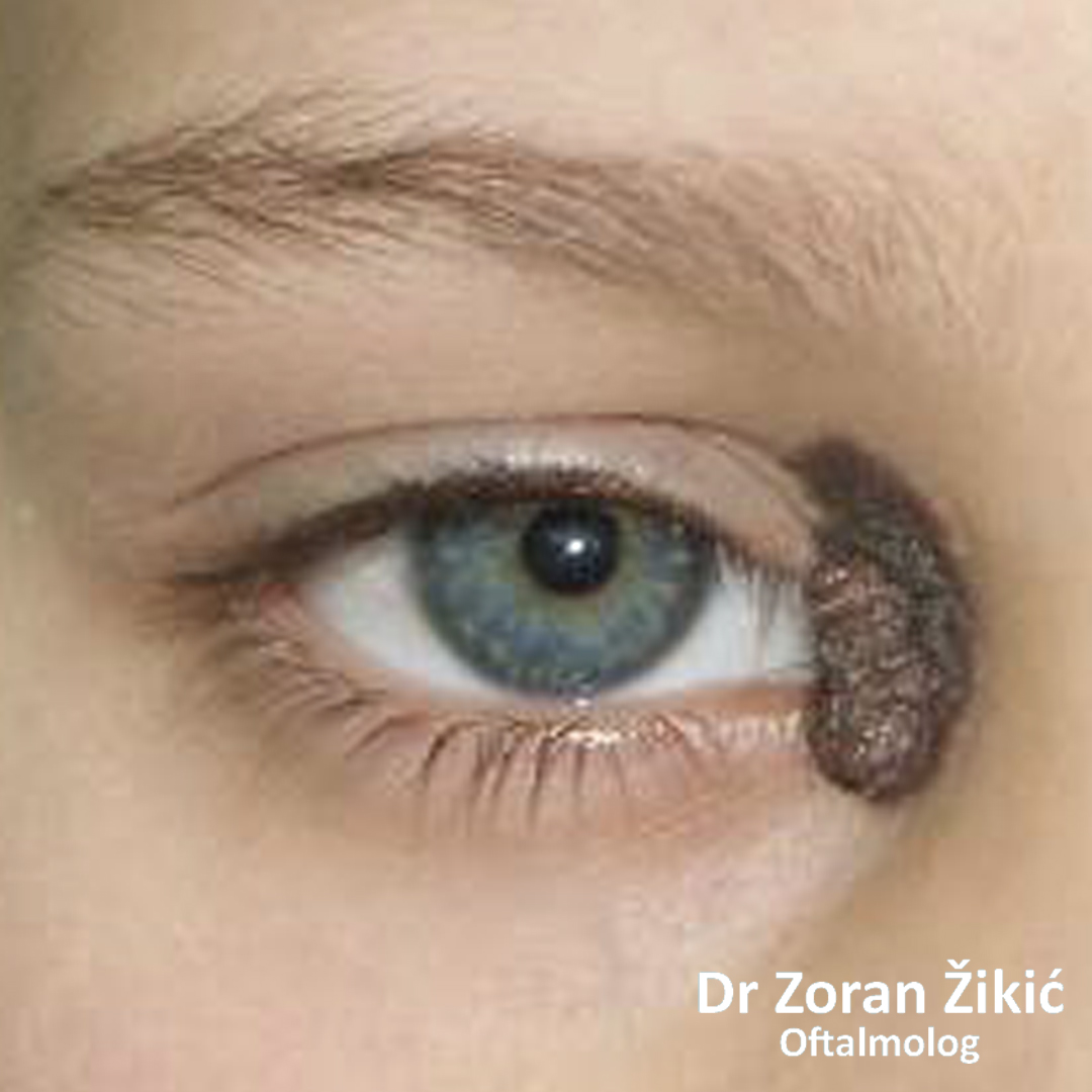
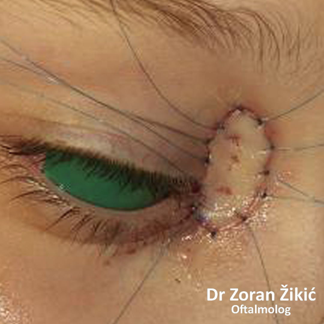
A big mole (nevus) of the inner angle, in a child.
Before and at the operation (the mole has been excised and replaced by a skin graft). The eye is protected by a green shield.
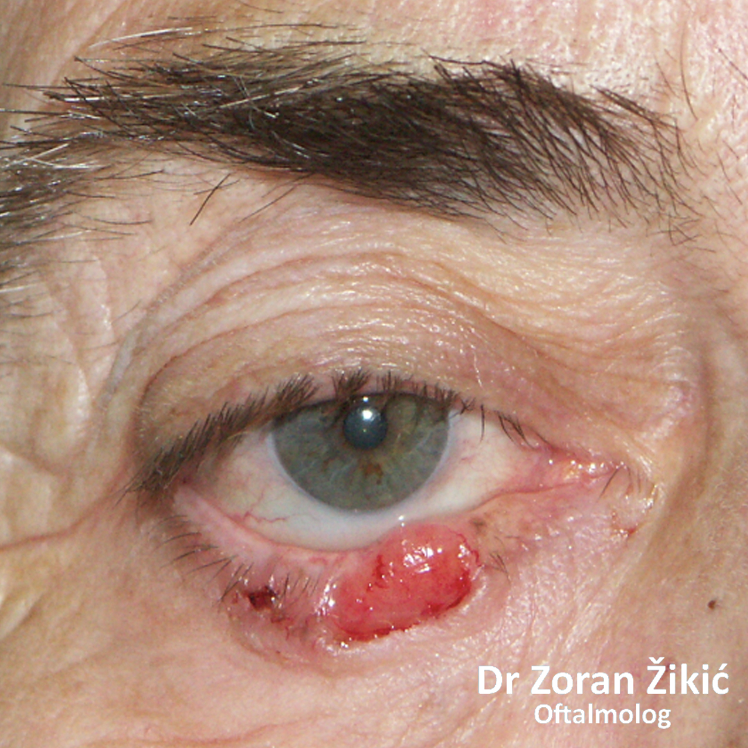
Tumour of the lower lid, causing it to evert.
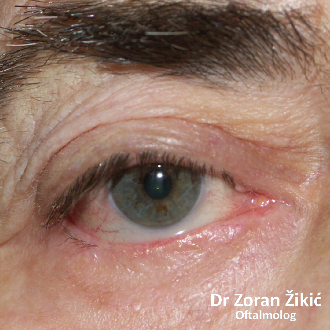
After surgery.
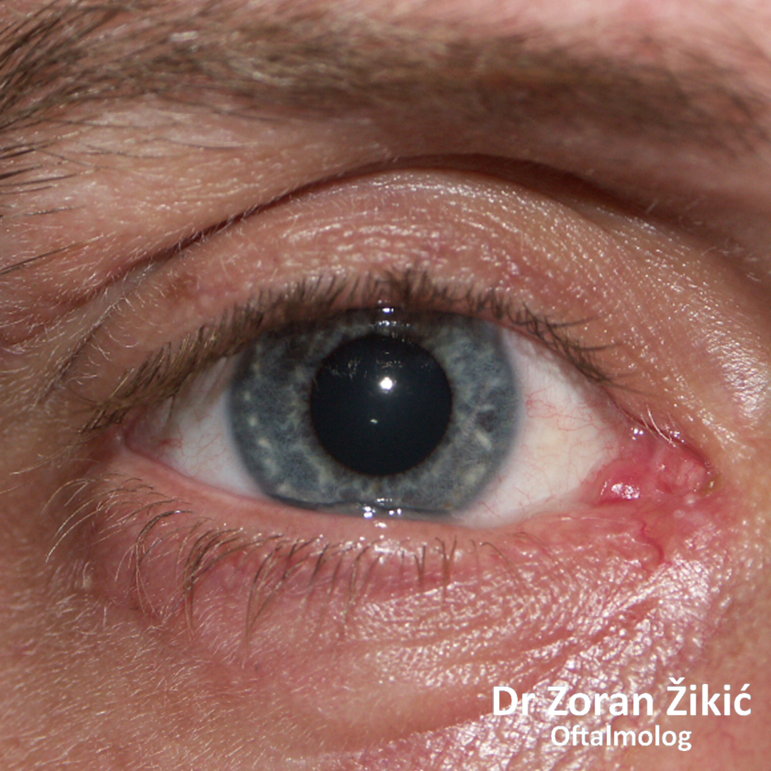
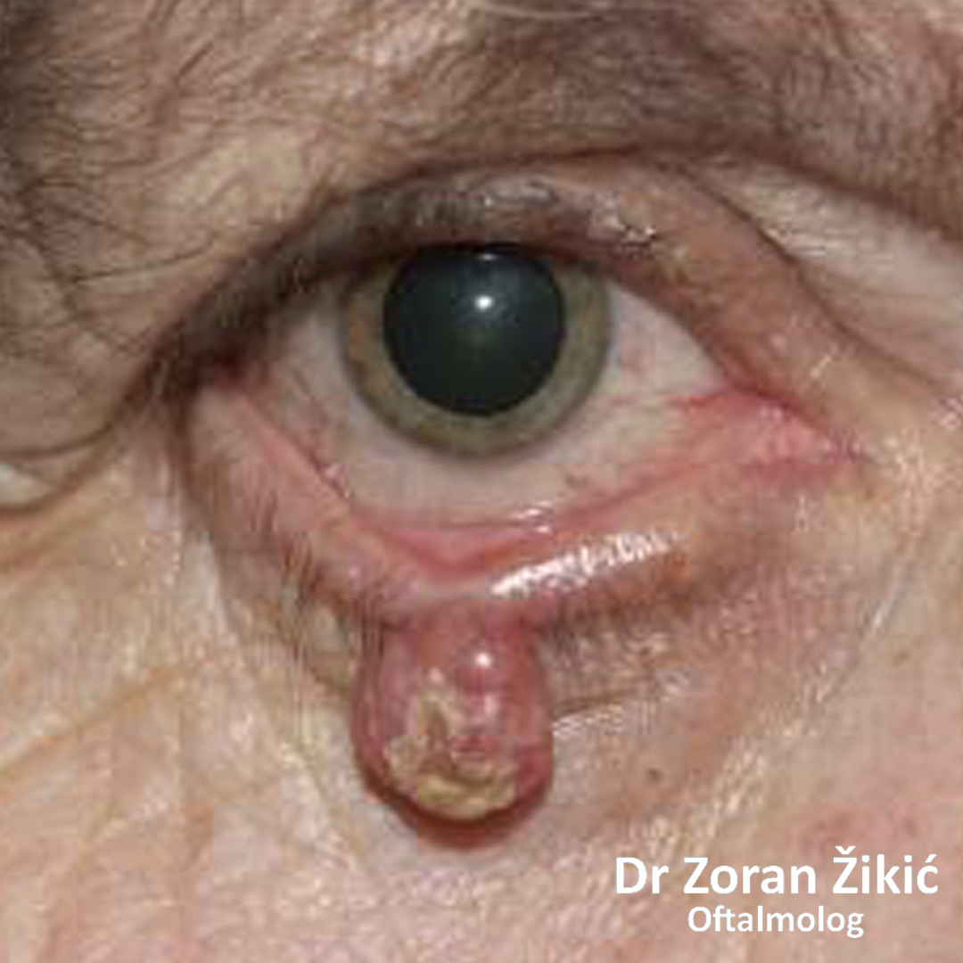
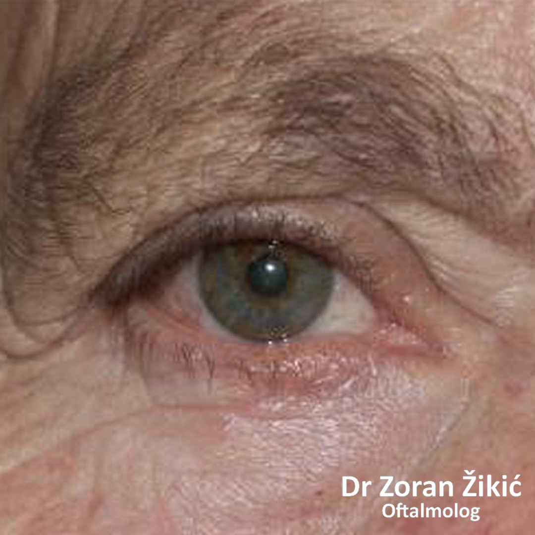
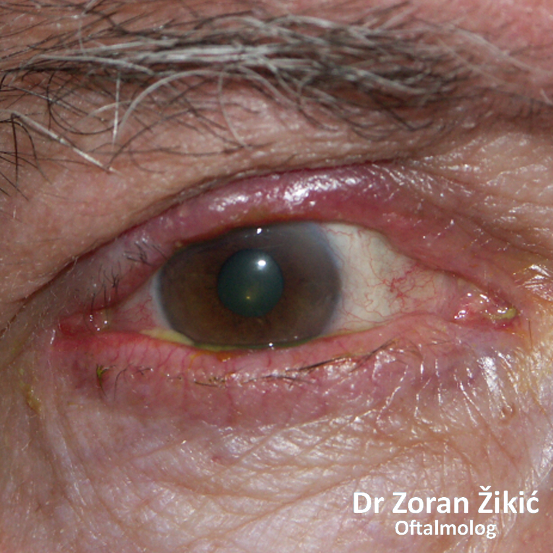
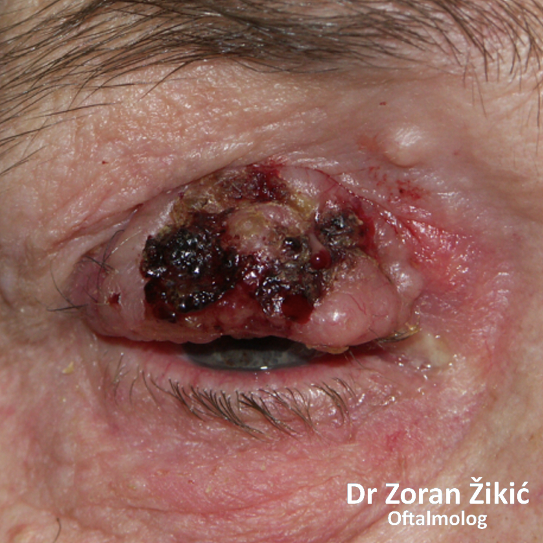
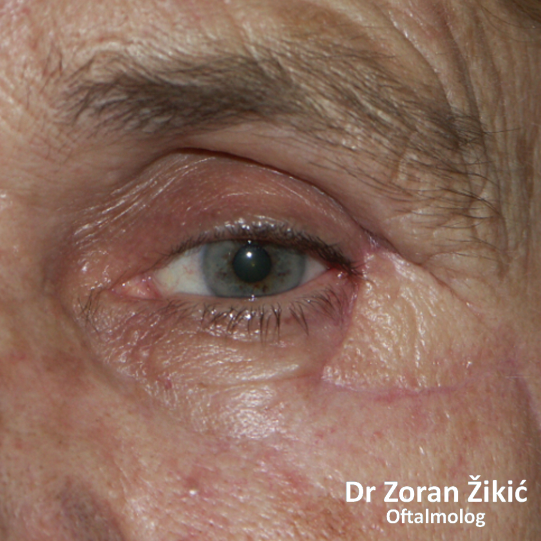
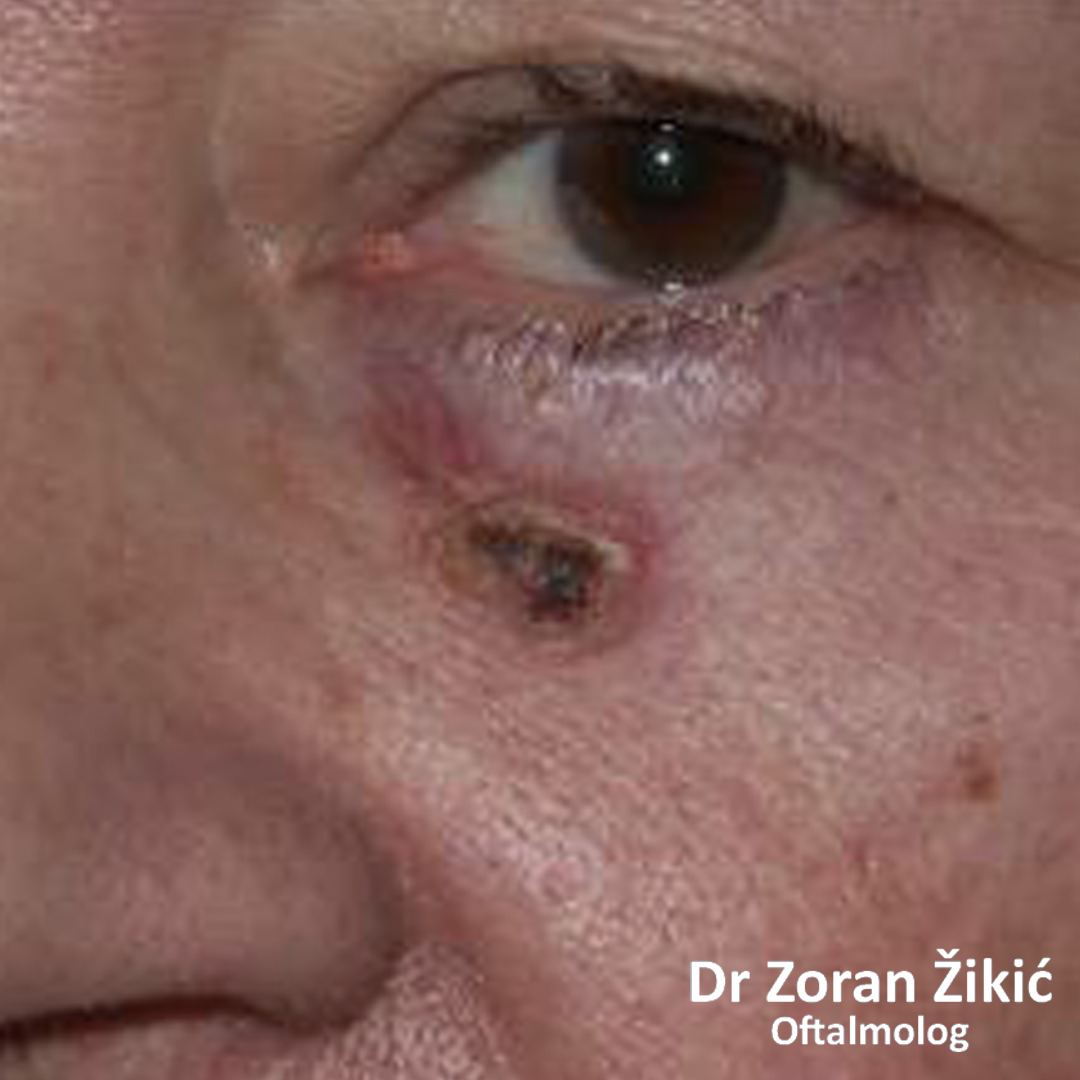
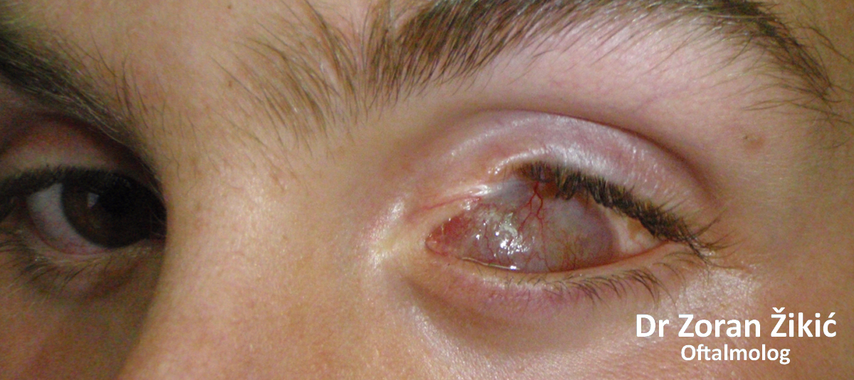
Tumour of the lower lid, as a non-healing ulcer..
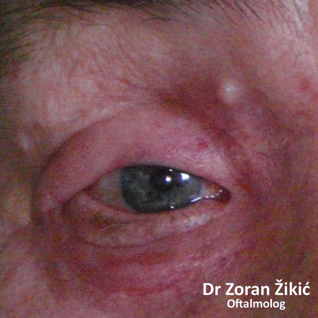
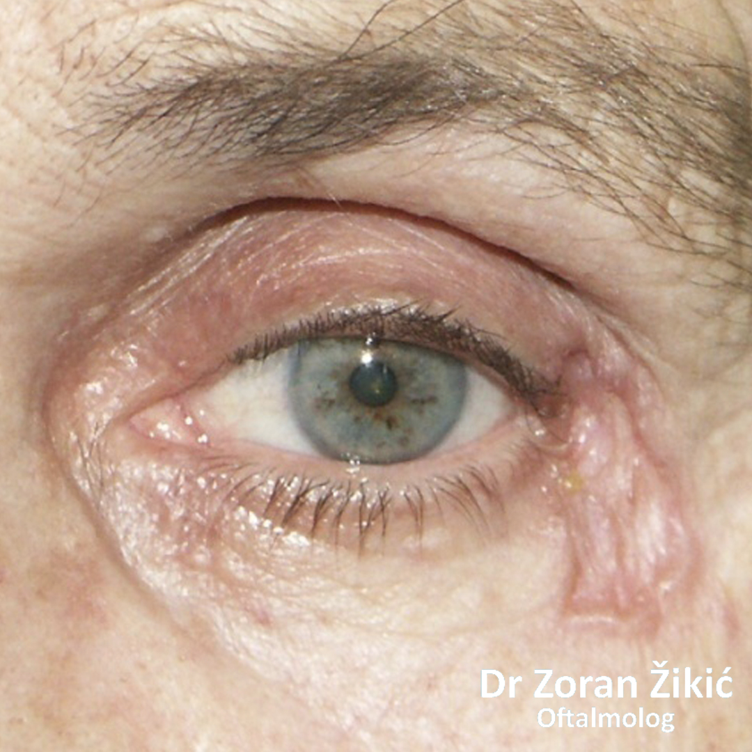
After surgery.
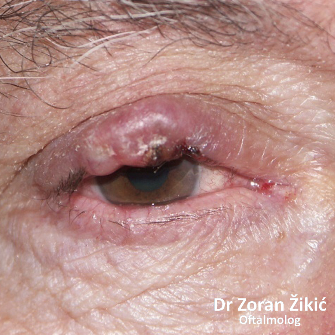
Tumour of the upper lid.
After surgery.
Tumour of the upper lid.
After surgery.
Tumour of the outer angle of the eyelids.
After surgery.
Tumour at the border of the lower lid and cheek.
After surgery.
CONGENITAL ANOMALIES OF THE EYELIDS
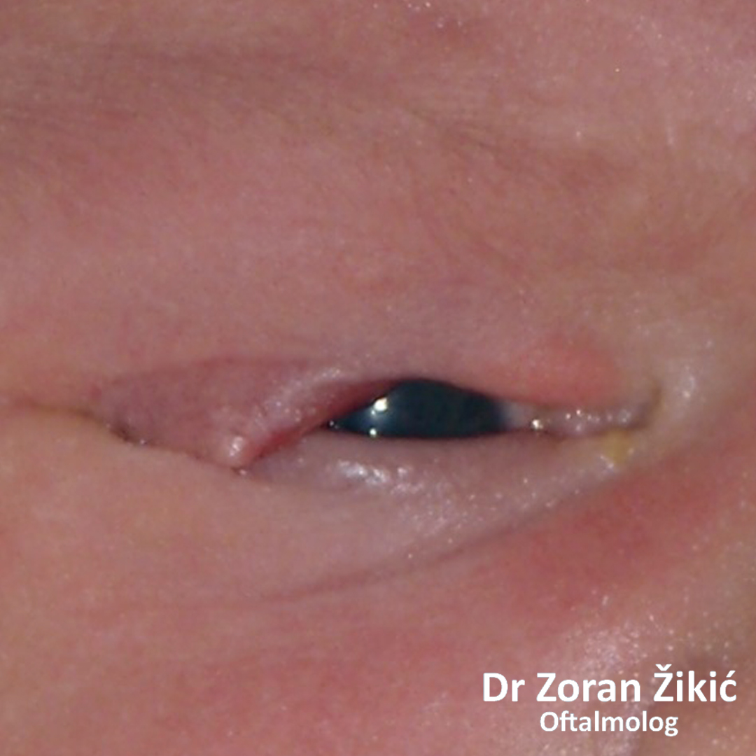
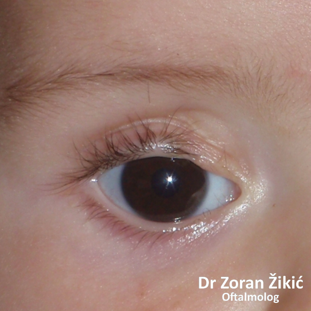
Congenital defect of the inner part of the upper lid in a premature infant.
8 months after reconstruction.
Congenital defect of pat of the upper lid (coloboma).
One year after reconstruction.
Congenital retraction of left lower lid.
After correction by retractor recession and hard pallate mucosa transplanatation.
RECONSTRUCTIVE SOCKET AND OCULAR PROSTHETIC SURGERY
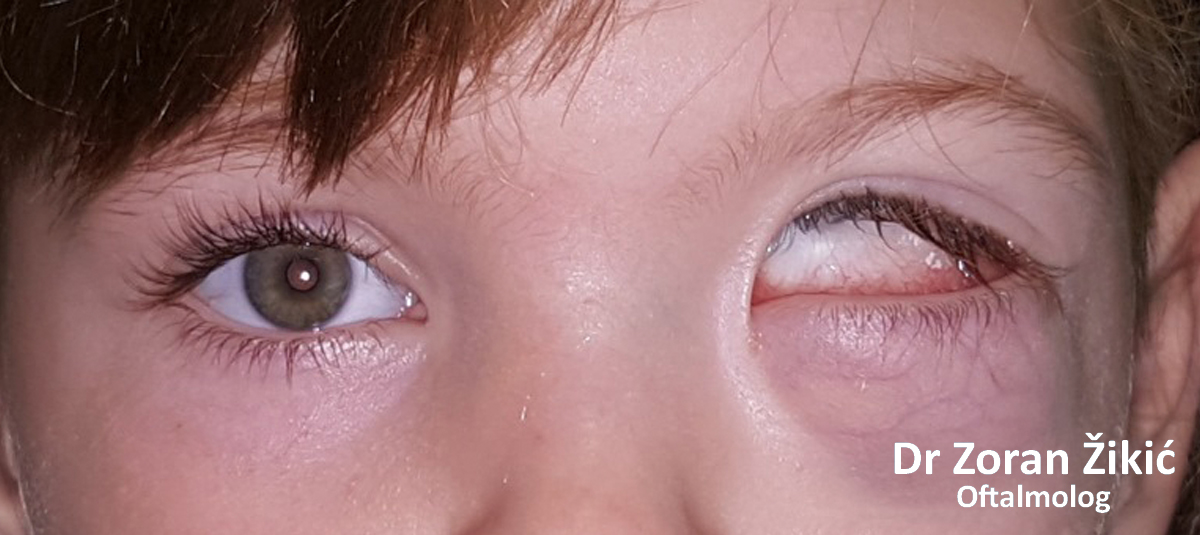
Congenital microphthalmos with a large orbital cyst.
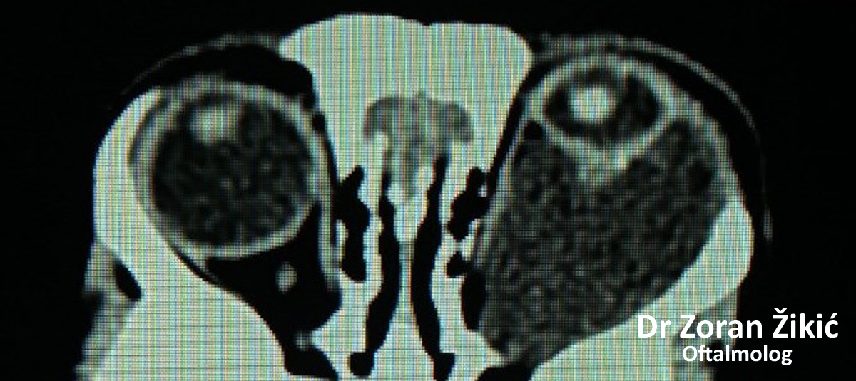
Computerized tomography (CT), which shows mechanical enlargement of the bones of the orbit.
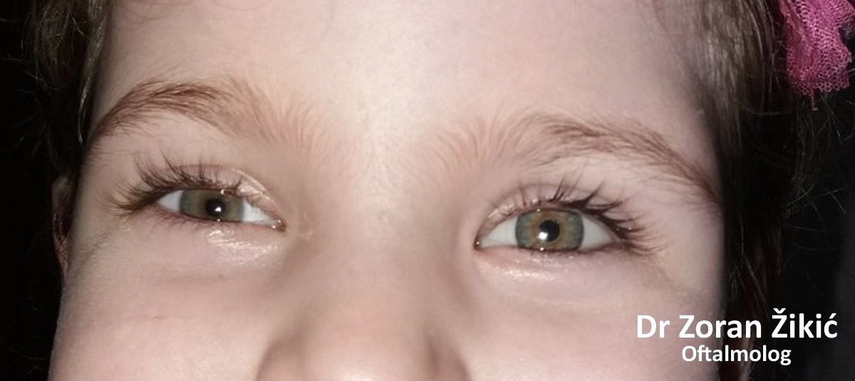
Situation after surgical removal of orbital cyst, enucleation of microphthalmic eye and dermis-fat transplant and prosthetic treatment.
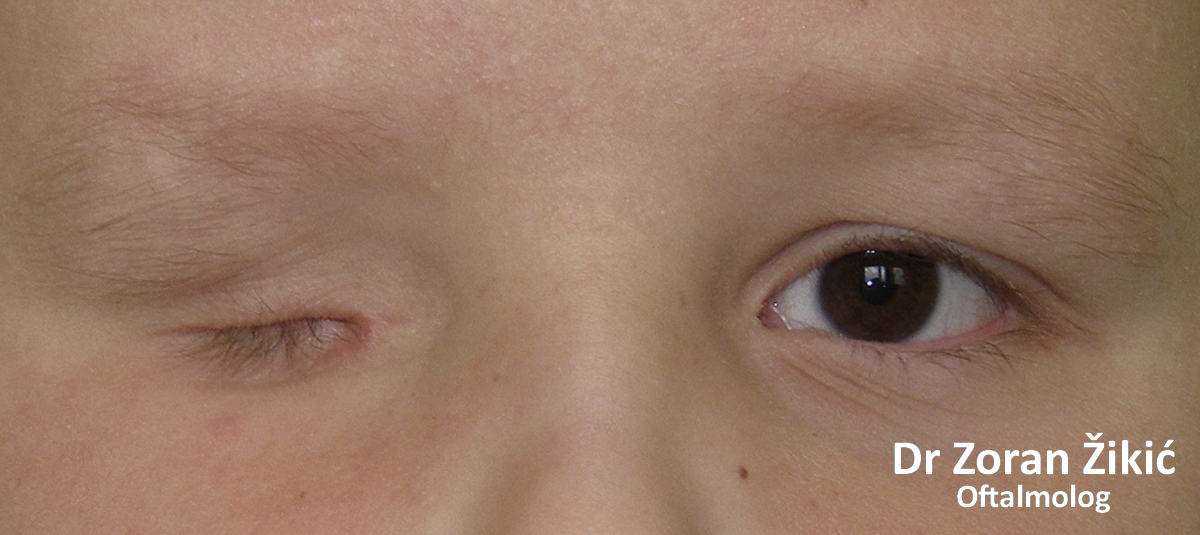
Congenital complete absence of the eye and underdevelopment of the orbit and eyelids (congenital microphtalmos).
After surgery and prosthetic treatment.
A large cyst in the orbit, after an injury of the left eye, which is blind.
After reconstructive surgery and prosthetic treatment.